

Foundation A
Question 1:
**Answer : B) Endochondral ossification**
**Explanation:**
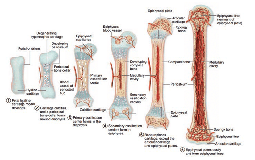
Endochondral ossification is the process of bone formation that takes place within a cartilage model. During fetal development, long bones such as the femur and humerus are formed through endochondral ossification. In this process, a hyaline cartilage model is gradually replaced by bone tissue. Intramembranous ossification, on the other hand, involves the direct conversion of mesenchymal tissue to bone, as seen in the formation of flat bones like the skull. Osteogenesis is a more general term for bone formation. Calcification refers to the deposition of calcium salts in tissue, which is part of the process of bone formation. Remodelling involves the continuous resorption and formation of bone tissue in response to mechanical and metabolic demands.
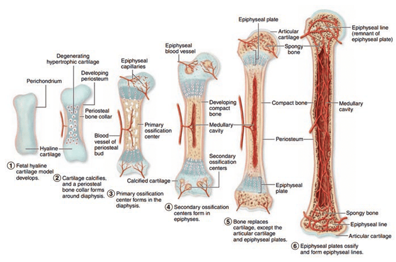
Stages of endochondral ossification:
Formation of Cartilage Model:
A cartilage model of the future bone, composed of hyaline cartilage, forms during early fetal development. This cartilage model is surrounded by a membrane called the perichondrium.
Growth of Cartilage Model:
Chondrocytes, the cells within the cartilage, proliferate and secrete extracellular matrix enzymes, which allows the cartilage model to grow in size.
Primary Ossification Centre Formation:
- Blood vessels migrate into the perichondrium releasing osteoblasts (bone forming cells). These osteoblasts convert the perichondrium (cartilaginous shaft) into a periosteum (bone).
- Blood vessels migrate from the periosteum & into the cartilage model, bringing osteoblasts into the centre of the diaphysis (the shaft of the bone). This area is now called the primary ossification centre.
- Chondrocytes in the primary ossification centre enlarge (hypertrophy) and then die, leaving cavities within the cartilage.
Bone Collar Formation:
Osteoblasts begin to secrete osteoid around the diaphysis of the cartilage model, forming a bone collar – compact bone surrounding the inner layer of the periosteum. This provides structural support.
Cavitation and Marrow Space Formation:
- The dying chondrocytes leave cavities that are invaded by periosteal bud containing blood vessels, nerves, red marrow elements, osteoblasts, and osteoclasts.
- Osteoclasts break down the calcified cartilage, and osteoblasts deposit new bone matrix, forming spongy/trabecular bone within the diaphysis.
Development of the Medullary Cavity:
As the primary ossification centre expands towards the epiphyses (ends of bone), osteoclasts break down the newly formed spongy bone in the diaphysis, creating the medullary (marrow) cavity.
Secondary Ossification Centres:
- Secondary ossification centres form in the epiphyses (the ends of the bone) after birth. Chondrocytes in these areas hypertrophy and die, and their spaces are invaded by blood vessels and osteoblasts, similar to the primary ossification centre.
- Osteoblasts replace the cartilage with spongy bone in the epiphyses.
Cartilage Replacement:
- Most of the cartilage is replaced by bone, except for two regions:
- Articular cartilage, which remains on the surfaces of the epiphyses to provide smooth surfaces for joint movement.
- The epiphyseal plates (growth plates), which are layers of cartilage that allow the bone to continue growing in length during childhood and adolescence.
Completion of Ossification:
Once growth in length is complete, the epiphyseal plates ossify and become the epiphyseal lines, marking the end of longitudinal bone growth.
Question 2:
**Answer: C) Ball-and-socket joint**
**Explanation:**
Ball-and-socket joints allow for a wide range of motion in multiple directions, as they permit movement in multiple planes, including rotation. Examples of ball-and-socket joints in the body include the shoulder (glenohumeral joint) and the hip (ischio-femoral joint).
Hinge joints, such as the elbow, permit movement in only one plane, like a door hinge.
Pivot joints, like the joint between the atlas and axis vertebrae, allow rotational movement around a central axis.
Saddle joints, found in the thumb, permit a variety of movements but not as extensive as ball-and-socket joints such as rotation.
Gliding aka planar joints allow for limited movement, typically in multiple directions within the same plane. The articulating surfaces of the bones in a gliding (or planar) joint are usually flat or slightly curved, allowing the bones to slide past each other. Examples of gliding joints include the intercarpal and intertarsal joints in the wrists and ankles like those between the carpal bones.

Question 3:
**Answer: B) Locus**
**Explanation:**
A locus is the specific physical location of a gene on a chromosome.
A gene is a segment of DNA that codes for a specific protein or RNA molecule.
A codon is a sequence of three nucleotides/bases that codes for a specific amino acid during protein synthesis.
An allele is a variant form of a gene that may result in different traits.
Homologs aka homologous chromosomes are pairs of chromosomes that carry similar genes but may carry different alleles, one from each parent.
An exon is a coding region of a gene that is transcribed into RNA and eventually translated into protein.

Question 4:
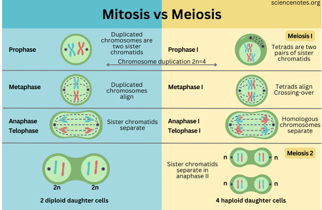
**Answer: B) Meiosis**
**Explanation:**
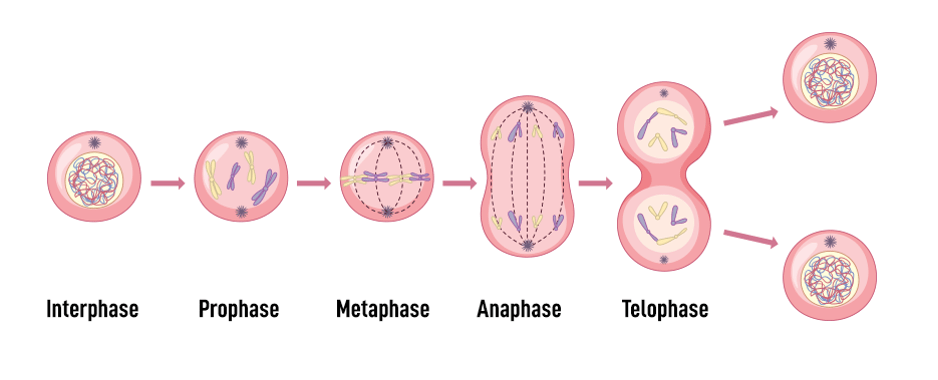
Meiosis is a cell division process that specifically involves the separation of homologous chromosomes, resulting in the formation of haploid cells (gametes). Mitosis, on the other hand, results in the formation of genetically identical diploid cells.
In meiosis, homologous chromosomes align next to each other (synapsis) during metaphase I and then separate during anaphase I. This is followed by a second division, where sister chromatids separate, leading to four haploid cells.
In mitosis, homologous chromosomes do not pair up. Instead, individual chromosomes align at the metaphase plate during metaphase. Sister chromatids then separate during anaphase, resulting in two genetically identical diploid daughter cells.
DNA replication is the process of copying DNA before cell division.
Transcription is the synthesis of mRNA from a DNA template. Translation is the process of synthesizing proteins using the information in mRNA.
Question 5:
**Answer: C) Sickle cell anaemia**
**Explanation:**
Sickle cell anaemia is a genetic disorder caused by a mutation in a single gene that affects the structure of beta chains in haemoglobin, leading to the formation of abnormal, sickle-shaped red blood cells.
Cystic fibrosis is caused by mutations in the CFTR gene resulting in thickened mucus and affects the respiratory and digestive systems.
Huntington’s disease is caused by a mutation in the HTT gene resulting in abnormal levels of the CAG codon. This affects the nervous system & causes neural damage & degeneration.
Down syndrome is caused by an extra copy of chromosome 21.
Haemophilia is an X-linked genetic disorder affecting blood clotting.
Question 6:
**Answer: B) Permitting ion exchange across synaptic clefts to propagate action potentials**
**Explanation:**
Neurotransmitters play a crucial role in synaptic signalling by binding to receptors on the postsynaptic neuron, which often leads to the opening or closing of ion channels. This ion exchange is essential for the propagation of action potentials and the transmission of signals between neurons.
A) Initiating kinase cascades for intracellular signalling Neurotransmitters primarily function by binding to receptors on the postsynaptic neuron to facilitate ion exchange and propagate action potentials, not by directly initiating kinase cascades for intracellular signalling. While some neurotransmitters can activate second messenger systems that may involve kinase cascades, this is not their primary role in synaptic signalling.
C) Transmitting signals along nerve fibres Neurotransmitters are involved in transmitting signals across synapses (the gaps between neurons), not along nerve fibres. The propagation of signals along nerve fibres (axons) is primarily achieved through the movement of action potentials.
D) Facilitating long-term changes in gene expression While neurotransmitters can influence processes that may lead to long-term changes in gene expression, such as through second messenger systems and transcription factors, their primary role in synaptic signalling is to permit ion exchange across synaptic clefts to propagate action potentials. Long-term gene expression changes are a downstream effect, not a direct role.
E) Modulating hormone release from endocrine cells Neurotransmitters primarily function within the nervous system to transmit signals between neurons. Modulating hormone release from endocrine cells is typically the function of neurohormones or direct neural control of endocrine glands, not the primary role of neurotransmitters in synaptic signalling.
Question 7:
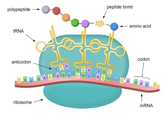
**Answer: B) Translation**
**Explanation:**
Translation is the process in which the sequence of codons on mRNA is decoded by ribosomes to synthesize a protein.
Transcription is the process of synthesizing mRNA from a DNA template.
Replication involves copying DNA during interphase stage of cell division.
Segregation refers to the separation of alleles during gamete formation.
Crossing over is the exchange of genetic material between homologous chromosomes during meiosis.
Question 8:
**Answer: C) Huntington’s disease**
**Explanation:**
Huntington’s disease is caused by an expansion of the CAG trinucleotide repeat in the HTT gene. This results in the accumulation of abnormal protein aggregates and the progressive degeneration of nerve cells, leading to motor and cognitive impairments.
Cystic fibrosis is caused by mutations in the CFTR gene.
Down syndrome is caused by an extra copy of chromosome 21.
Haemophilia is an X-linked genetic disorder affecting blood clotting.
Turner syndrome involves a missing or partially missing X chromosome in females.
Question 9:
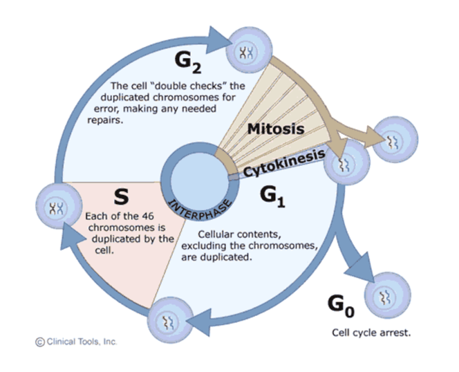
**Answer: B) S phase**
**Explanation:**
DNA replication occurs during the S (synthesis) phase of the cell cycle. In this phase, DNA is duplicated to ensure that each daughter cell receives a complete copy of the genetic material.
The G1 phase is the first gap phase where the cell grows and synthesizes proteins and organelles to prepare for DNA replication.
G2 is the second gap phase where the cell continues to grow and prepares for mitosis, increasing the volume of cytoplasm and ensuring all proteins and organelles are ready for cell division.
M phase is the mitotic phase when cell division occurs.
Although interphase is correct, it is not the best answer since interphase includes G1, S & G2 phase. Only S phase of interphase actually involves DNA replication.
Question 10:
**Answer: A) Mitosis**
**Explanation:**
Mitosis is a process of cell division that produces two genetically identical daughter cells, each with the same number of chromosomes as the parent cell (diploid cell).
Meiosis, on the other hand, involves two rounds of division and results in the formation of four haploid cells with genetic variation & half the number of chromosomes as the parent cell.
Binary fission is a form of asexual reproduction in prokaryotes.
Budding is a type of asexual reproduction seen in some organisms.
Question 11:
**Answer: C) Hinge joint**
**Explanation:**
A hinge joint permits movement in only one plane, resembling the motion of a door hinge. Examples include the elbow joint and the knee joint.
Ball-and-socket joints allow movement in multiple directions such as rotation, circumduction, adduction & abduction, medial & lateral rotation etc., like the shoulder and hip.
Pivot joints allow rotation around a central axis, as seen in the neck (between C1 & C2 vertebrae).
Saddle joints allow various movements but not rotation.
Gliding joints permit sliding or gliding movements.
Question 12:
**Answer: E) Modification and packaging of molecules**
**Explanation:**
The Golgi apparatus is responsible for modifying, sorting, and packaging proteins and lipids for transport within or outside the cell. It consists of flattened membranous sacs called cisternae.
Energy production occurs in the mitochondria.
DNA replication takes place in the cell nucleus.
Protein synthesis occurs on ribosomes.
Lipid synthesis occurs in the smooth endoplasmic reticulum.
Question 13:
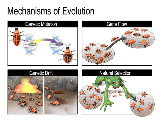
**Answer: B) Gene flow**
**Explanation:**
Gene flow refers to the movement of alleles from one population to another through migration, leading to a change in allele frequencies within both populations.
Genetic drift is the random change in allele frequencies due to chance events.
Natural selection is the process by which organisms with advantageous traits survive and reproduce more successfully.
Mutation introduces new alleles into a population.
Speciation is the process by which new species arise.
Question 14:
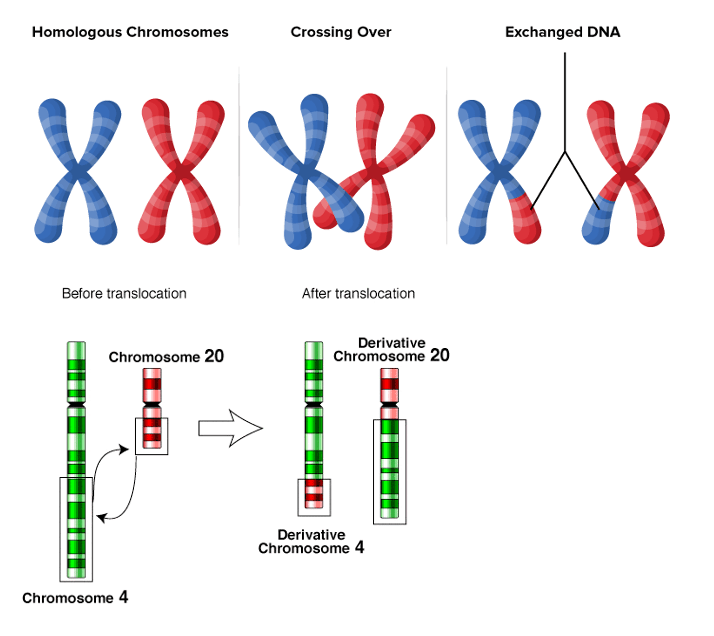
**Answer: A) Crossing over**
**Explanation:**
Crossing over is the process during meiosis where genetic material is exchanged between non-sister chromatids of homologous chromosomes. This results in genetic recombination and contributes to genetic diversity among offspring.
Recombination refers to the mixing of alleles from different sources.
Segregation is the separation of alleles during gamete formation.
Replication is the copying of DNA in the S phase of interphase.
Translocation involves the movement of genetic material between different chromosomes.
Question 15:
**Answer: D) M phase**
**Explanation:**
The M (mitotic) phase is characterized by the division of the cell nucleus (which occurs in prophase stage of mitosis) and the distribution of replicated chromosomes into two daughter cells (anaphase stage of mitosis).
G1, S, and G2 phases collectively make up interphase, during which the cell prepares for division.
Cytokinesis, which follows mitosis, involves the division of the cytoplasm and organelles into the daughter cells.
Question 16:
**Answer: C) Mitochondria**
**Explanation:**
Mitochondria are the sub-cellular organelles responsible for producing ATP through oxidative phosphorylation. They are often referred to as the “powerhouses” of the cell due to their role in energy production. The process takes place in the inner mitochondrial membrane.
The endoplasmic reticulum is involved in protein synthesis (rough endoplasmic reticulum) and lipid metabolism (smooth endoplasmic reticulum).
The Golgi apparatus modifies, sorts, and packages molecules.
Many biochemical processes occur in the cytoplasm such as substrate level phosphorylation (e.g. glycolysis).
The nucleus contains genetic information.
Question 17:
**Answer: C) Apoptosis**
**Explanation:**
Apoptosis is a controlled and programmed form of cell death that is essential for maintaining tissue homeostasis, proper development, and removing damaged or unnecessary cells (such as death of cells in the tail of the embryo, death of leukocytes that have already been used in an immune response to prevent autoimmunity & the death of cells containing damaged or mutated DNA). It is characterized by cell shrinkage, chromatin condensation, and the formation of apoptotic bodies.
Necrosis is a form of cell death caused by irreversible cellular injury or disease. Necrosis causes loss of membrane integrity & inflammation.
Autophagy is the degradation/recycling of cellular components.
Mitosis is the process of cell division that results in two genetically identical daughter cells.
Anoikis is a form of cell death in which cells lose their anchorage to the extra-cellular membrane. This death is vital in preventing metastasis in cancer.
Question 18:
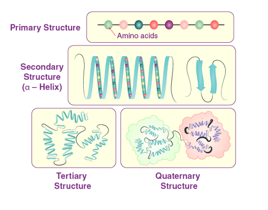
**Answer: A) Primary structure**
**Explanation:**
The primary structure of a protein refers to the linear sequence of amino acids in its polypeptide chain.
Secondary structure involves the local folding patterns, such as alpha helices and beta sheets which are stabilised by hydrogen bonds between the backbone amide and carbonyl groups.
Tertiary structure is the overall three-dimensional arrangement of a single protein molecule. This is caused by various interactions, including hydrogen bonds, ionic bonds, hydrophobic interactions, and disulphide bridges between R groups of different amino acids.
Quaternary structure refers to the arrangement of multiple protein subunits in a larger protein complex &/or the addition of a prosthetic group such as heme in haemoglobin.
Oligomeric structure describes the association of a few protein subunits in a complex, which is a specific type of quaternary structure.

Question 19:
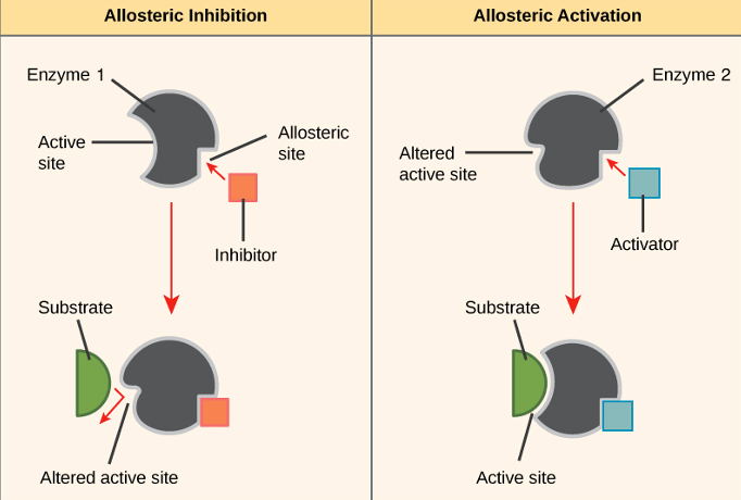
**Answer: A) Allosteric regulation**
**Explanation:**
Allosteric regulation involves the binding of a regulatory molecule at a site other than the active site of an enzyme, leading to a conformational change that affects the enzyme’s activity. Allosteric regulation is a type of non-competitive inhibition but can involve both activation and inhibition, while non-competitive inhibition specifically refers to inhibition. Therefore, allosteric regulation is a better answer since the question does not refer to inhibition only.
Competitive inhibition occurs when a molecule competes with the substrate for the active site.
Non-competitive inhibition involves binding at a different site, but it still affects the active site’s function.
Feedback regulation involves a product inhibiting an enzyme in its biosynthetic pathway.
Cooperativity refers to one substrate molecule influencing the binding of additional substrate molecules (such as the binding of oxygen to haemoglobin which causes a sigmoid-shaped curve to form).
Question 20:
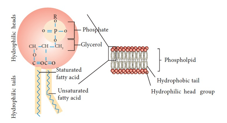
**Answer: D) Lipids**
**Explanation:**
Lipids are biomolecules characterised by their hydrophobic nature meaning they repel water. This hydrophobic property is crucial for their role as major components of the phospholipid bilayer of cellular membranes. The hydrophobic tails of phospholipids face inward, away from water, while the hydrophilic heads face outward, interacting with the aqueous environment. This arrangement forms a stable barrier that separates the cell from its surroundings and regulates the passage of substances in and out of the cell.
Amino acids are the building blocks of proteins being predominantly hydrophilic.
Nucleic acids, such as DNA and RNA, carry genetic information & are hydrophilic.
Saccharides are sugars and carbohydrates which are hydrophilic.
Ions are charged particles involved in cellular signalling and electrolyte balance.
Question 21:
**Answer: B) S phase**
**Explanation:**
The S (synthesis) phase of the cell cycle is characterized by active DNA replication. During this phase, the DNA is duplicated to prepare for cell division.
The G1 and G2 phases are gap phases where the cell grows and prepares for DNA replication (G1) and cell division (G2).
G0 phase is a quiescent stage where cells exit the cell cycle and do not divide; it is also the stage where cells can specialise and perform their functions.
Interphase includes G1, S, and G2 phases collectively thus S phase is a better answer.
Question 22:
**Answer: A) Genetic drift**
**Explanation:**
Genetic drift is the process that involves random changes in allele frequency due to chance events. This can lead to significant changes in a population’s genetic makeup over time, especially in small populations.
Gene flow refers to the transfer of alleles or genes from one population to another.
Natural selection involves changes in allele frequencies due to differential survival and reproduction of individuals based on their traits.
Mutation is the process by which new alleles are created through changes in DNA sequences.
Speciation is the formation of new and distinct species during evolution.
Question 23:
**Answer: E) Differentiation**
**Explanation:**
Differentiation is the process by which cells become specialised for specific functions within an organism. For example, stem cells in the colonic crypt may differentiate into villous cells & have absorptive functions, or they may differentiate into colonic crypt cells & have secretory functions.
Dedifferentiation refers to the reverse process, where specialised cells revert to a less specialised state.
Proliferation is the rapid division of cells.
Apoptosis is programmed cell death.
Cellular fusion involves the merging of two cells.
Question 24:
**Answer: A) Rough endoplasmic reticulum**
**Explanation:**
The rough endoplasmic reticulum (RER) is responsible for protein synthesis, containing ribosomes that synthesise proteins. The rough ER is studded with ribosomes, giving it a “rough” appearance, while the smooth ER lacks ribosomes and is involved in lipid metabolism and detoxification.
The Golgi apparatus modifies and packages proteins in order to be exported out of the cell.
Mitochondria produce ATP.
Vesicles are membrane-bound organelles involved in transport within the cell. They can also fuse with the cell membrane to release molecules out of the cell e.g. in fusing with the pre-synaptic membrane of neurones to release neurotransmitters.
Question 25:
**Answer: D) Nucleic acids**
**Explanation:**
Nucleic acids are biomolecules primarily composed of a sequence of nucleotide bases. Each nucleotide consists of three components:
- A sugar (deoxyribose in DNA or ribose in RNA)
- A phosphate group
- A nitrogenous base (adenine, thymine/uracil, cytosine, and guanine in DNA; adenine, uracil, cytosine, and guanine in RNA)
These nucleotides are linked together by phosphodiester bonds to form DNA or RNA molecules, which carry genetic information and play crucial roles in protein synthesis and other cellular functions.
Amino acids are the building blocks of proteins. They are composed of a carbon skeleton, amino & carboxyl groups.
Carbohydrates are composed of sugars and serve as energy sources or structural components.
Triglycerides are lipids composed of glycerol and three fatty acids, serving as energy storage molecules.
Monoglycerides are lipids composed of glycerol and one fatty acid, also involved in lipid metabolism.
Question 26:
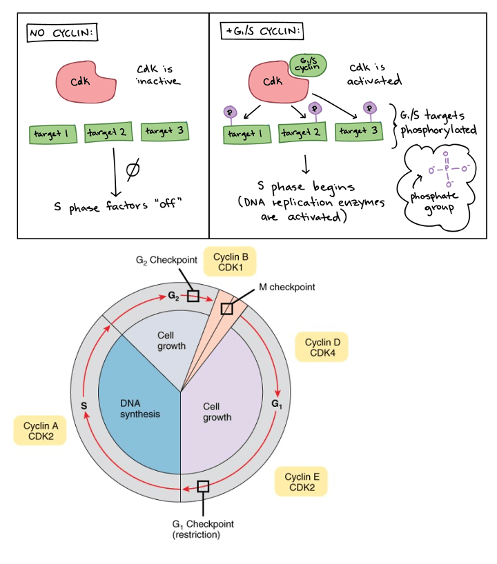
**Answer: C) They regulate the progression through cell cycle checkpoints.**
**Explanation:**
Cyclins and cyclin-dependent kinases (CDKs) are key regulators of the cell cycle. Cyclins bind to CDKs, activating them, and together they help control the transition from one phase of the cell cycle to another, especially at critical checkpoints. This ensures that the cell progresses through the cell cycle only when conditions are appropriate. DNA replication is facilitated by DNA polymerases. Cellular dedifferentiation is not a primary function of cyclins and CDKs. Cellular fusion involves the merging of two cells.
Question 27:
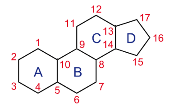
**Answer: A) Steroid hormones**
**Explanation:**
Steroid hormones, which are hydrophobic in nature & are lipid soluble, can easily pass through cell membranes. They act on target cells after being transported through the bloodstream and binding to intracellular receptors. Steroid hormones have a distinct characteristic of which they are all derived from cholesterol & have the ABCD appearance.
Peptide hormones, amino acid-derived hormones, and eicosanoids usually act through extracellular receptors located on cell membranes.
Thyroid hormones are transported through the bloodstream, are lipid-soluble, and can act on intracellular receptors. Unlike steroid hormones, thyroid hormones are derived from amino acids, specifically tyrosine not cholesterol therefore option A is the correct answer.
Question 28:
**Answer: D) Quaternary structure**
**Explanation:**
Quaternary structure refers to the spatial arrangement of multiple protein subunits in a larger protein complex &/or the addition of a prosthetic group. Primary structure is the sequence of amino acids. Secondary structure involves local folding patterns, like alpha helices and beta sheets. Tertiary structure is the overall three-dimensional arrangement of a single protein molecule. Oligomeric structure describes the association of a few protein subunits.
Question 29:
**Answer: A) Primary active transport**
**Explanation:**
Primary active transport is the process that involves the movement of solutes against their concentration gradient with the help of membrane proteins, typically utilising energy in the form of ATP. This energy is directly used to transport molecules such as ions (e.g., sodium, potassium) across the cell membrane against their concentration gradient.
Simple diffusion is the movement of molecules from an area of higher concentration to an area of lower concentration down its concentration gradient, without the need for membrane proteins or energy input.
Osmosis is the movement of water across a selectively permeable membrane from an area of lower solute concentration to an area of higher solute concentration.
Secondary active transport involves the coupled transport of molecules across the membrane, where the movement of one molecule down its concentration gradient provides energy for the movement of another molecule against its gradient. This method does not require ATP.
Facilitated diffusion is the passive transport of molecules across the membrane facilitated by transport proteins, but it occurs along the concentration gradient and does not require energy input.
Question 30:
**Answer: D) Enabling the passage of ions between cells**
**Explanation:**
Ligand-gated ion channels play a critical role in cell-cell communication by enabling the passage of ions between cells upon ligand binding. This allows for rapid electrical and chemical signalling between adjacent cells.
Transmitting signals along nerve fibres is typically mediated by voltage-gated ion channels, which respond to changes in membrane potential rather than ligand binding.
Facilitating long-term changes in gene expression usually involves intracellular receptors and signalling pathways rather than ion channels.
Initiating kinase cascades for intracellular signalling is often mediated by receptor tyrosine kinases or G-protein coupled receptors, not ligand-gated ion channels.
Modulating hormone release from endocrine cells is usually controlled by exocytosis and cellular signalling mechanisms specific to hormone secretion.
Question 31:
**Answer: B) Paracrine signalling**
**Explanation:**
Paracrine signalling involves the release of molecules into the extracellular space to affect nearby cells, even if they are not in direct physical contact with the secreting cell. Juxtacrine signalling, while involving direct contact, is specifically about signals transmitted through physical contact rather than released molecules.
Autocrine signalling involves cells responding to their own molecules.
Endocrine signalling affects distant target cells through the bloodstream.
Synaptic signalling occurs between nerve cells.
Juxtacrine signalling involves direct communication between adjacent cells.
Question 32:
**Answer: D) Gliding joint**
**Explanation:**
Gliding joints are found between the flat surfaces of bones, allowing limited movement in multiple directions. Examples of gliding joints include the sacroiliac joint, sternoclavicular joint & joints between carpal & tarsal bones.
Ball-and-socket joints allow a wide range of motion, as seen in the shoulder and hip.
Pivot joints permit rotation around a central axis, like the neck.
Hinge joints allow movement in one plane, such as the elbow.
Saddle joints allow various movements, as seen in the thumb.
Question 33:
**Answer: B) Golgi apparatus**
**Explanation:**
The Golgi apparatus is responsible for modifying, sorting, and packaging proteins and lipids for transport within or outside the cell.
The endoplasmic reticulum is involved in protein synthesis and lipid metabolism.
Mitochondria produce ATP.
Vesicles are involved in transport.
The nucleus contains genetic information.
Question 34:
**Answer: A) Ligand-gated receptor**
**Explanation:**
Ligand-gated receptors, also known as ionotropic receptors, are commonly found in the plasma membrane. These receptors respond to specific signalling molecules (ligands) by opening ion channels, allowing the passage of ions across the membrane. This rapid ion flow through the channels influences the electrical properties of the cell membrane and can trigger various cellular responses.
Tyrosine kinase receptors are membrane-bound receptors that phosphorylate tyrosine residues on intracellular proteins in response to ligand binding.
G-protein coupled receptors (GPCRs) activate intracellular signalling pathways through G proteins upon ligand binding activating (Gs)/ inhibiting (Gi) adenylate cyclase or activating phospholipase C (Gq).
Nuclear receptors are intracellular receptors that bind to ligands and regulate gene expression.
Voltage-gated receptors are ion channels that open or close in response to changes in membrane potential (voltage).
Question 35:
**Answer: C) Endocrine signalling**
**Explanation:**
Endocrine signalling refers to the process of cell-cell communication where signalling molecules (hormones) are released into the bloodstream by endocrine glands or cells. These hormones travel through the bloodstream to reach distant target cells or tissues, where they exert their effects by binding to specific receptors.
Autocrine signalling involves cells responding to their own molecules.
Paracrine signalling targets nearby cells.
Synaptic signalling occurs at specialised nerve cell junctions.
Juxtacrine signalling involves direct communication between adjacent cells.
Question 36:
**Answer: D) Changes in gene transcription**
**Explanation:**
Nuclear receptors are involved in cell-cell communication by binding to hydrophobic signalling molecules, such as steroid hormones, and initiating changes in gene transcription. Steroid hormones or thyroid hormones, which are lipid-soluble, diffuse through the cell membrane into the cytoplasm or nucleus where they bind to intracellular nuclear receptors. Upon binding, the receptor-hormone complex undergoes a conformational change and forms a dimer structure that acts as a transcription factor. This transcription factor complex then binds to specific DNA sequences (response elements) in the nucleus, stimulating or inhibiting gene transcription. These changes in gene expression lead to long-term cellular responses mediated by the synthesis of new proteins or other regulatory molecules. Nuclear receptors do not activate G-protein pathways, facilitate cell adhesion, initiate kinase cascades, or facilitate rapid ion exchange.
Question 37:
**Answer: A) Transcription**
**Explanation:**
Transcription is the process of converting a DNA sequence into an RNA molecule. This RNA, known as messenger RNA (mRNA), carries the genetic information from the DNA to the ribosomes for protein synthesis during translation. Replication involves copying DNA during cell division. Segregation refers to the separation of alleles during gamete formation. Differentiation is the process of specialisation of cells.
Question 38:
**Answer: B) Meiosis I**
**Explanation:**
Meiosis I is the process of cell division that involves the separation of homologous chromosomes, resulting in the formation of haploid cells. Meiosis II follows and involves the separation of sister chromatids. Both meiosis I & meiosis II involved the formation of haploid cells however separation of homologous chromosomes only occurs in meiosis I.
Mitosis results in the formation of genetically identical diploid cells.
Binary fission is a form of asexual reproduction in prokaryotes.
Budding is a type of asexual reproduction seen in some organisms.
Question 39:
**Answer: E) Eicosanoids**
**Explanation:**
These are signalling molecules derived from arachidonic acid, a fatty acid found in cell membranes. Eicosanoids are produced locally at the site of action in response to stimuli such as injury or inflammation. They include prostaglandins, thromboxanes, and leukotrienes, which exert their effects on nearby cells and tissues. Eicosanoids are not typically released into the bloodstream to act on distant cells but instead act locally where they are synthesised.
Steroid hormones are lipid-soluble molecules derived from cholesterol. They diffuse across cell membranes and bind to intracellular receptors, exerting their effects by altering gene transcription. However, they are transported through the bloodstream to reach target tissues and organs throughout the body.
Peptide hormones are composed of amino acids and are synthesised in the endocrine glands. They are released into the bloodstream and act on target cells that possess specific receptors for the hormone.
Amino acid derived hormones include hormones like adrenaline and thyroid hormones, which are synthesised from amino acids and tyrosine. They are released into the bloodstream and act on distant target cells.
Question 40:
**Answer: E) Gap junction-mediated communication**
**Explanation:**
Gap junctions are specialised intercellular connections found between cells, including cardiac muscle cells (cardiomyocytes), at structures called intercalated discs. These junctions allow for direct electrical and metabolic communication between adjacent cells such as calcium & sodium flow by forming channels known as connexons. These channels permit the passage of ions and small molecules, facilitating rapid depolarization and coordinated contraction of the heart muscle.
Autocrine signalling involves cells responding to signalling molecules that they themselves produce.
Paracrine signalling involves the release of signalling molecules into the extracellular space to affect nearby cells.
Desmosome junction-mediated communication involves strong adhesive junctions that anchor cells together and provide mechanical stability, but they do not facilitate direct communication or ion passage between cells.
Tight junction-mediated communication forms a barrier between cells to regulate the passage of ions and molecules, primarily maintaining cellular polarity and barrier function rather than enabling communication.
Question 41:
**Answer: A) Activation of neighbouring cells by signalling molecules**
**Explanation:**
The bystander effect occurs when signalling molecules released by one cell activate or influence nearby cells. This phenomenon highlights the ability of certain signalling molecules (such as paracrine factors or cytokines) to diffuse through the extracellular space and affect adjacent cells, even if those cells were not the primary target of the initial signal. The activation of neighbouring cells can amplify or propagate cellular responses within a tissue or organ, contributing to coordinated physiological processes or responses to stimuli.
Passive diffusion of signalling molecules between cells describes the physical process of signalling molecules moving through the extracellular space from one cell to another.
Inhibition of cellular response to signalling molecules refers to mechanisms where cells may reduce their sensitivity or responsiveness to specific signalling molecules over time.
Activation of G-proteins by ligands describes a mechanism where ligands bind to G-protein coupled receptors (GPCRs) on the cell surface, leading to intracellular signalling cascades.
Opening of ligand-gated channels in adjacent cells refers to the direct effect where ligand binding to receptors on one cell opens ion channels on that cell’s membrane, influencing its electrical properties.

Question 42:
**Answer: A) Autocrine signalling**
**Explanation:**
Autocrine signalling involves cells releasing signalling molecules that affect their own function such as prostaglandins, interleukin-1 & epidermal growth factor. Paracrine signalling affects nearby cells. Endocrine signalling targets distant cells through the bloodstream. Synaptic signalling occurs between nerve cells. Juxtacrine signalling involves direct communication between adjacent cells.

Question 43:
**Answer: D) Nuclear receptor**
**Explanation:**
Nuclear receptors are involved in the binding of hydrophobic signalling molecules such as steroid hormones. Upon ligand binding, nuclear receptors translocate to the nucleus and directly affect gene transcription. Ligand-gated receptors, tyrosine kinase receptors, GPCRs, and ion channel receptors are not primarily responsible for changes in gene transcription.

Question 44:
**Answer: E) To initiate cascades for intracellular signalling**
**Explanation:**
Receptor tyrosine kinases (RTKs) play a crucial role in cell-cell communication by initiating kinase cascades for intracellular signalling upon ligand binding. When a hormone binds to tyrosine kinase receptors e.g., insulin, this causes the receptors to dimerise, phosphorylating each other. As a result, this triggers an intracellular cascade such as the translocation of GLUT4 to the cell membrane enabling glucose to be absorbed into the cell. RTKs are not directly involved in regulating ion channels or G-protein signalling. Ligand-gated channels are opened by ligand binding. Cell adhesion is mediated by different types of receptors.

Question 45:
**Answer: D) Control of digestion and gastrointestinal motility**
**Explanation:**
The enteric nervous system is a complex network of nerves that controls digestion and gastrointestinal motility, including processes like peristalsis and secretion. The ENS operates largely independently of the central nervous system (CNS), often referred to as the “second brain.” However, it is also influenced by the CNS. The parasympathetic nervous system (part of the autonomic nervous system) can up-regulate ENS activity, promoting digestive processes (rest & digest), while the sympathetic nervous system can down-regulate ENS activity, inhibiting digestion (fight or flight).The other options do not accurately describe the primary role of the enteric nervous system.
Question 46:
**Answer: D) ATP stores energy in its high-energy phosphate bonds.**
**Explanation:**
ATP molecules consist of adenine (a nitrogenous base), ribose (a sugar), and three phosphate groups. The bonds between these phosphate groups are high-energy bonds, meaning they store a significant amount of potential energy. When ATP is hydrolysed by the enzyme ATPase, the bond between the last two phosphate groups (known as the terminal phosphate) is cleaved, releasing energy. This reaction produces adenosine diphosphate (ADP) and an inorganic phosphate molecule (Pi). The released energy from ATP hydrolysis is used to drive various cellular processes that require energy, such as: muscle contraction, active transport & intracellular process such as DNA replication.

Question 47:
**Answer: B) Glycolysis**
**Explanation:**
Glycolysis is the metabolic pathway that breaks down glucose (a 6-carbon sugar) into two molecules of pyruvate (a 3-carbon compound).This process occurs in the cytoplasm of the cell and generates a small amount of ATP (2 molecules per glucose molecule) and NADH (reduced form of NAD+).
After glycolysis, pyruvate is transported into the mitochondria. In the mitochondrial matrix, pyruvate undergoes oxidation and decarboxylation in the link reaction. This results in the formation of acetyl CoA, which is a 2-carbon compound bound to coenzyme A (CoA).
Acetyl CoA enters the Krebs cycle, a series of biochemical reactions that occur in the mitochondrial matrix. During the Krebs cycle, acetyl CoA is oxidized & decarboxylated to carbon dioxide, generating ATP, NADH, and FADH2 (reduced forms of NAD+ and FAD).
NADH and FADH2 produced in glycolysis, the link reaction, and the Krebs cycle donate electrons to the electron transport chain (ETC), located in the inner mitochondrial membrane. As electrons move through the ETC, they release energy that is used to pump protons (H^+ ions) across the membrane, creating an electrochemical gradient. ATP synthase then utilises the energy from the proton gradient to phosphorylate ADP to ATP, a process known as oxidative phosphorylation. This process produces a large amount of ATP (approximately 28-34 molecules per glucose molecule).
Gluconeogenesis is the synthesis of glucose from non-carbohydrate sources such as amino acids, lactate, and glycerol.
Question 48:
**Answer: C) Computed Tomography (CT)**
**Explanation:**
Computed Tomography (CT) is a medical imaging technique that uses X-rays to create detailed cross-sectional images (also called slices) of the body. These images provide clear views of bones, internal organs, and soft tissues, making CT scans particularly useful for detecting various medical conditions, including cancers. CT scans are widely used in clinical practice for diagnosing and monitoring conditions such as tumours, fractures, infections, and vascular diseases.
Ultrasounds use sound waves to create images of internal organs. It is useful for imaging soft tissues and monitoring fetal development but is not as helpful in visualising bones or tumours.
MRIs utilise strong magnetic fields and radio waves to produce detailed images of organs and tissues. MRI is excellent for soft tissue imaging and does not involve ionising radiation, but it is less commonly used for bone imaging compared to CT.
PET scans involve injecting a radioactive tracer into the body to detect metabolic activity in tissues. PET scans are used in oncology to assess cancer spread and treatment response but do not provide detailed anatomical images like CT.
Fluoroscopy uses continuous X-rays to create real-time images of internal structures. It is helpful for guiding procedures but does not produce detailed cross-sectional images like CT.
Question 49:
**Answer: C) Germinal centres**
**Explanation:**
Germinal centres are specialised regions within lymph nodes where B cells undergo intense proliferation, somatic hypermutation, and affinity maturation in response to antigens. B cells that encounter specific antigens migrate to germinal centres within lymph nodes. Within germinal centres, B cells undergo somatic hypermutation, a process where their antibody genes undergo random mutations. This diversifies the antibody repertoire, potentially improving the affinity of antibodies for the antigen. B cells that produce antibodies with higher affinity for the antigen are selectively expanded through a process called affinity maturation. Some B cells differentiate into memory B cells, which provide long-term immunity upon re-exposure to the antigen, while others differentiate into plasma cells that secrete large quantities of antibodies into the bloodstream.
The thymus is an organ where T cells mature, not where B cells undergo proliferation and differentiation.
Antibody-producing centres is not as specific to the structured regions within lymph nodes where B cell processes occur.
Memory cells are a type of B cell that provides immunological memory after initial exposure to an antigen.
Plasma cells are differentiated B cells that produce and secrete antibodies.
Question 50:
**Answer: E) Humoral immunity**
**Explanation:**
Humoral immunity involves the production of antibodies by B lymphocytes. It provides rapid defence against familiar pathogens and is a component of adaptive immunity.
Innate immunity is the first line of defence against infections.
Cellular immunity involves T lymphocytes.
Passive immunity is the transfer of antibodies from one individual to another e.g., from mother to fetus.
Question 51:
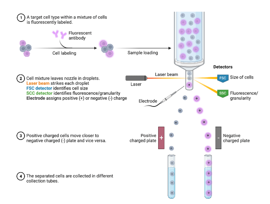
**Answer: D) To identify surface antigens on cells and quantitatively analyse cellular characteristics**
**Explanation:**
Fluorescence-activated cell sorting (FACS) analysis, or flow cytometry, is a powerful technique used in cell biology and immunology to analyse and sort cells based on various characteristics, primarily focusing on surface antigens.
- FACS can detect and quantify specific surface antigens on individual cells by labelling them with fluorescently-tagged antibodies. This helps researchers identify different cell types or subsets within a heterogeneous population.
- FACS allows for the quantitative analysis of cellular characteristics such as size, granularity (complexity), and fluorescence intensity of labelled antibodies. This provides insights into cell populations and their functional states.
- FACS can sort cells based on their fluorescence profiles, allowing isolation of specific cell populations for further study or experimentation.
Analysing DNA sequences is conducted using techniques like PCR (Polymerase Chain Reaction) or DNA sequencing methods, not FACS.
Protein secondary structures are typically studied using techniques like X-ray crystallography or NMR spectroscopy, not FACS.
Studying the distribution of organelles is done using electron microscopy.
ATP levels are typically measured using biochemical assays or luminescence-based techniques, not FACS.
Question 52:
**Answer: C) Erythropoietin **
**Explanation:**
Erythropoietin (EPO) is a key growth factor/cytokine involved in the development of red blood cells (erythropoiesis). EPO is primarily produced by the kidneys in response to low oxygen levels in the blood (hypoxia). It stimulates the bone marrow to produce red blood cells from hematopoietic stem cells.
Glucagon & insulin hormones are involved in regulating glucose metabolism.
Epinephrine & norepinephrine are involved in the “fight or flight” response.
Oestrogen & progesterone are involved in reproductive functions and have
Testosterone is involved in male reproductive functions, while FSH stimulates the growth of ovarian follicles in females.
Question 53:
**Answer: A) Erythrocytes**
**Explanation:**
Red blood cells (erythrocytes) are specialised for oxygen transport from the lungs to body tissues and carbon dioxide transport from tissues to the lungs. Erythrocytes contain haemoglobin, a protein that binds to oxygen molecules in the lungs. Oxygenated haemoglobin (oxyhaemoglobin) is then transported by erythrocytes through the bloodstream to tissues where oxygen is released. Erythrocytes also carry carbon dioxide from body tissues back to the lungs. Carbon dioxide is either dissolved in the plasma or converted into bicarbonate ions within erythrocytes to facilitate transport.
White blood cells (leukocytes) are involved in the immune response.
Platelets (thrombocytes) are important for blood clotting.
Reticulocytes are immature blood cells that eventually mature into erythrocytes.
Question 54:
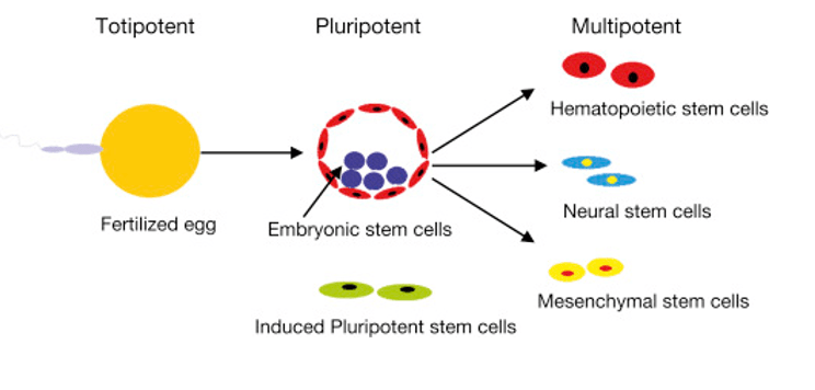
**Answer: D) Totipotent**
**Explanation:**
Totipotent cells are undifferentiated cells with the ability to differentiate into any cell type. They have the ability to differentiate into all cell types of the body, including both embryonic and extraembryonic tissues (such as placental cells). Totipotent cells are found only in the very early stages of embryonic development, such as the cells of the zygote and the cells produced by the first few divisions of the fertilized egg blastomeres & morula.
Pluripotent cells, on the other hand, are cells that can differentiate into all cell types derived from the three germ layers (ectoderm, mesoderm, and endoderm) of the embryo, but not into extraembryonic tissues like the placenta.
Multipotent cells can differentiate into a limited number of cell types within a particular lineage or tissue. They are more specialised than pluripotent cells. Examples include hematopoietic stem cells (which can differentiate into various blood cell types) and mesenchymal stem cells.
Progenitor cells are more differentiated than stem cells but less specialized than fully differentiated cells. They can differentiate into a limited number of cell types related to their specific lineage or tissue.
Unipotent cells can only differentiate into one type of cell, typically their own type or a closely related type.
Question 55:
**Answer: D) Cardiac muscle**
**Explanation:**
Cardiac muscle is characterised by branching fibres that are interconnected by intercalated discs, which allow synchronized contractions of the heart. Skeletal muscle is responsible for voluntary movements. Smooth muscle is found in internal organs, in not branched & is spindle shaped. Striated muscle includes skeletal and cardiac muscle. Voluntary muscle is synonymous with skeletal muscle.

Question 56:
**Answer: A) Oxidative phosphorylation**
**Explanation:**
Oxidative phosphorylation refers to the process of generating ATP using the energy released during the electron transport chain and the movement of protons (H⁺ ions) across a membrane. In the inner mitochondrial membrane (or the plasma membrane in prokaryotes), electrons derived from NADH and FADH₂ are passed through a series of electron transporter complexes in the electron transport chain. As electrons move through the electron transport chain, this releases energy which pumps protons (H⁺ ions) across the membrane from the mitochondrial matrix to the intermembrane space creating an electrochemical gradient. The electrochemical gradient allows protons to move back across the membrane through ATP synthase, a protein complex embedded in the membrane (by facilitated diffusion). This movement of protons powers ATP synthase to catalyse the phosphorylation of ADP to ATP, utilising the energy released from the proton gradient.
Glycolysis is a metabolic pathway that breaks down glucose to pyruvate, generating a small amount of ATP through substrate-level phosphorylation in the cytoplasm.
Substrate level phosphorylation involves the direct transfer of a phosphate group from a phosphorylated substrate to ADP to form ATP, occurring during glycolysis and the citric acid cycle.
Gluconeogenesis is the synthesis of glucose from non-carbohydrate precursors such as amino acids and glycerol.
Citric acid cycle also known as the Krebs cycle, is a series of chemical reactions that oxidize acetyl-CoA to produce ATP, NADH, FADH₂, and CO₂ in the mitochondrial matrix, involving substrate level phosphorylation.
Question 57:
**Answer: B) Gluconeogenesis**
**Explanation:**
Gluconeogenesis is the process of producing glucose from non-carbohydrate precursors such as glycerol, amino acids, and lactate. Glycerol, derived from the breakdown of fats (triglycerides), serves as a precursor for gluconeogenesis. Other substrates include amino acids from proteins and lactate from anaerobic glycolysis. Gluconeogenesis primarily occurs in the liver and to a lesser extent in the kidneys. It is an energy-demanding process that requires ATP and GTP. Gluconeogenesis is important for maintaining blood glucose levels during fasting or starvation periods when glucose availability from dietary sources is limited. It also provides glucose for tissues that cannot use fatty acids as an energy source, such as red blood cells and certain parts of the brain.
Glycolysis is the metabolic pathway that breaks down glucose to pyruvate in the cytoplasm, producing ATP and NADH.
Glycogenosis involves the conversion of glucose into glycogen via insulin to be stored in liver, muscle & adipose tissue.
Beta-oxidation is the process of breaking down fatty acids into acetyl-CoA molecules for energy production through the citric acid cycle.
Glycogenolysis is the breakdown of glycogen into glucose-1-phosphate, which can be further converted to glucose-6-phosphate and then to free glucose.
Question 58:
**Answer: E) Oculomotor nerve (III)**
**Explanation:**
The oculomotor nerve (cranial nerve III) is responsible for controlling most of the muscles that move the eye (except for the superior oblique and lateral rectus muscles, which are controlled by cranial nerves IV and VI, respectively). The oculomotor nerve innervates the medial rectus, inferior rectus, superior rectus, and inferior oblique muscles of the eye, which are responsible for moving the eye in different directions. The oculomotor nerve also controls the sphincter pupillae muscle in the iris, which constricts the pupil in response to light (pupillary light reflex) or during accommodation for near vision (near response).
Olfactory nerve (I) is responsible for the sense of smell.
Optic nerve (II) is responsible for vision, transmitting visual information from the retina to the brain.
Trigeminal nerve (V) is responsible for sensation in the face, including the corneal reflex but not for eye movements or pupil constriction.
Trochlear nerve (IV) is responsible for innervating the superior oblique muscle of the eye, which helps in downward and inward eye movements (intorsion and depression).
Question 59:
**Answer: D) GPCRs activate adenylate cyclase or phospholipase C to generate second messengers.**
**Explanation:**
GPCRs are integral membrane proteins that play a pivotal role in transmitting signals from extracellular ligands to intracellular signalling pathways. Upon binding of specific ligands (such as hormones or neurotransmitters like catecholamines) to the extracellular domain of the GPCR, conformational changes occur within the receptor. These changes activate intracellular G proteins associated with the GPCR. During Gs, alpha subunit activates adenylate cyclase which converts ATP into cyclic AMP (cAMP). Cyclic AMP then serves as a second messenger that activates protein kinase A (PKA), initiating a cascade of phosphorylation events that regulate various cellular processes such as metabolism, gene expression, and cell growth.
GPCRs do not serve as ion channels, regulate gene expression through tyrosine kinase activation, or facilitate direct cell-cell adhesion.
Question 60:
**Answer: C) Receptor tyrosine kinase (RTK)**
**Explanation:**
The insulin receptor is a type of receptor tyrosine kinase (RTK). RTKs are cell surface receptors that have intrinsic enzymatic activity. When insulin binds to the insulin receptor, it activates the receptor’s tyrosine kinase activity, leading to autophosphorylation of tyrosine residues on the receptor itself. This phosphorylation event then triggers a cascade of intracellular signalling pathways that ultimately regulate glucose uptake by cells, primarily through the translocation of glucose transporters (such as GLUT4) to the cell membrane.
GPCRs primarily activate intracellular signalling pathways through G proteins, not tyrosine kinase activity. They are not associated with insulin signalling or glucose uptake.
Ligand-gated ion channels open in response to specific ligand binding to allow ion passage across the membrane. They are not involved in insulin-mediated glucose uptake mechanisms.
Mechanoreceptors respond to mechanical stimuli like touch or pressure.
Voltage-gated receptors open and close in response to changes in membrane potential.
Question 61:
**Answer: C) Polymerase chain reaction (PCR)**
**Explanation:**
Polymerase chain reaction (PCR) is a laboratory technique used to amplify a specific DNA sequence. It involves repeated cycles of denaturation, annealing of primers, and DNA synthesis by DNA polymerase. This process leads to the generation of many copies of the targeted DNA segment.
Next-generation sequencing is a technique used to sequence millions of DNA fragments simultaneously, but it does not involve primer-based amplification of a specific DNA sequence.
DNA sequencing determines the order of nucleotides in a DNA molecule but does not necessarily involve amplification using primers.
DNA transcription is the process of synthesising RNA from a DNA template and is not related to amplification of a specific DNA sequence using primers.
Sanger sequencing is a method of DNA sequencing that involves chain termination and gel electrophoresis to determine the sequence of nucleotides, but it does not involve primer-based amplification of DNA.

Question 62:
**Answer: C) To identify individuals based on variations in short tandem repeat (STR) sequences**
**Explanation:**
STR profiling, also known as DNA fingerprinting, is used in forensic science to identify individuals based on variations in specific short tandem repeat (STR) sequences within their DNA. This technique relies on the unique patterns of repeated sequences to distinguish individuals. The options mentioning protein structure, nucleotide sequencing, metabolic pathways, and gene expression are not accurate descriptions of STR profiling.

Question 63:
**Answer: D) Sanger sequencing relies on chain termination and gel electrophoresis, while NeXT Gen sequencing uses real-time detection of nucleotide incorporation. **
**Explanation:**
Sanger sequencing relies on the termination of DNA synthesis using chain-terminating dideoxynucleotides (ddNTPs) and subsequent separation of the terminated fragments by gel electrophoresis. The sequence is read based on the positions of the terminated fragments. Next-Generation (NeXT Gen) DNA sequencing methods, on the other hand, do not use chain termination and gel electrophoresis. Instead, these methods involve massively parallel sequencing technologies that detect the incorporation of nucleotides in real-time as DNA polymerase adds them to the growing DNA strand. This allows for simultaneous sequencing of millions of DNA fragments in a highly parallel manner.
Both Sanger sequencing and Next-Generation sequencing methods can use fluorescently labelled nucleotides for detection, and radioactive labels are not commonly used in modern sequencing technologies.
Both Sanger sequencing and Next-Generation sequencing methods can use fluorescently labelled nucleotides for detection, and radioactive labels are not commonly used in modern sequencing technologies.
Both Sanger sequencing and Next-Generation sequencing methods require relatively small amounts of DNA template for sequencing, though Next-Generation methods often require higher total amounts due to the parallel nature of the sequencing process.
Question 64:
**Answer: A) Co-dominance**
**Explanation:**
Co-dominance is the phenomenon in Mendelian genetics where both alleles of a heterozygous individual are fully expressed. This means that neither allele is dominant or recessive to the other, and both contribute equally to the phenotype. An example of co-dominance is the AB blood group system in humans, where individuals with the genotype AB have both A and B antigens expressed on their red blood cells.
In incomplete dominance, the heterozygous phenotype is an intermediate or blended expression of the two alleles. An example is the pink flowers produced from crossing red and white flowered plants.
Epistasis refers to the interaction between different genes where one gene masks or modifies the phenotypic expression of another gene.
Penetrance refers to the proportion of individuals carrying a particular genotype who express the associated phenotype to any degree.
Polygenic inheritance occurs when a trait is influenced by multiple genes, each contributing small effects to the phenotype.

Question 65:
**Answer: C) X-linked inheritance**
**Explanation:**
X-linked inheritance involves traits determined by genes located on the X chromosome. These traits can be dominant or recessive and are often expressed differently in males and females due to the presence of one or two X chromosomes. The Y chromosome primarily determines male sex characteristics and does not typically carry genes that contribute to other traits.
Autosomal dominant inheritance involves a trait that is expressed when only one copy of the dominant allele is present on an autosome (non-sex chromosome).
Autosomal recessive inheritance occurs when an individual inherits two copies of a recessive allele on an autosome to express the trait.
Y-linked inheritance involves traits that are inherited solely through genes on the Y chromosome, which are primarily responsible for male sex determination and related characteristics.
Mitochondrial inheritance involves the transmission of traits through genes located in the mitochondria, which are inherited maternally causing all born offspring to be affected.
Question 66:
**Answer: C) Magnetic Resonance Imaging (MRI)**
**Explanation:**
Magnetic Resonance Imaging (MRI) is a medical imaging technique that provides detailed images of soft tissues, such as the brain, muscles, joints, and internal organs. It uses strong magnetic fields and radio waves to generate images based on the water content and density of tissues. MRI is particularly useful for detecting abnormalities and diseases in soft tissues that may not be well visualised with other imaging techniques like X-rays (option A), which are better suited for visualising bones and dense structures.
X-ray is more suited for visualising bones and dense tissues because they are absorbed by dense structures and show up as white on X-ray images.
Ultrasound uses sound waves to create images of organs and tissues. It is commonly used for imaging soft tissues and organs in real-time and is safe and non-invasive.
CT scans use X-rays to create cross-sectional images of the body. While it provides detailed images of both soft tissues and bones, it involves exposure to ionising radiation making option C the best answer.
PET scans are used to detect metabolic activity and function in tissues. They involve the injection of a radioactive tracer and are used to detect diseases such as cancer and monitor treatment response.
Question 67:
**Answer: D) Ca2+**
**Explanation:**
Calcium ions (Ca2+) are released from the sarcoplasmic reticulum in response to an action potential. Ca2+ binds to troponin C, causing a conformational change that allows myosin heads to bind to actin filaments. This initiates the cross-bridge cycle and muscle contraction. The other ions do not play a direct role in this process.

Question 68:
**Answer: B) Smooth muscle**
**Explanation:**
Smooth muscle is responsible for involuntary movements of internal organs. It is found in the walls of hollow organs such as the stomach, intestines, bladder, and blood vessels. Smooth muscle contracts and relaxes involuntarily, without conscious control, to facilitate functions like digestion, peristalsis, and regulation of blood flow.
Skeletal muscle is responsible for voluntary movements and is attached to bones by tendons. It is under conscious control and allows for movements such as walking, running, and lifting weights.
Cardiac muscle is an involuntary muscle, however is only found in the heart and is responsible for pumping blood throughout the body.
Striated muscle includes both skeletal and cardiac muscle, characterised by striped or striated appearance due to the arrangement of contractile proteins (actin and myosin).
“Voluntary muscle” is not a standard term used in anatomy or physiology. Skeletal muscle is typically referred to as voluntary muscle due to its conscious control, but it does not accurately describe the type of muscle responsible for involuntary movements of internal organs.
Question 69:
**Answer: C) Blood vessels grow into the perichondrium. Cells in the perichondrium differentiate into osteoblasts and begin forming bone around the edge of the cartilage shaft known as the periosteum.**
**Explanation:**
During stage 2 of endochondral ossification, blood vessels invade the perichondrium, which triggers the differentiation of cells within the perichondrium into osteoblasts. These osteoblasts begin forming a bone collar around the cartilage shaft, which becomes the periosteum. This process is crucial for providing a support structure for the developing bone and marks the transition from cartilage to bone.
Option A is incorrect because this describes the first stage of endochondral ossification, where chondrocytes enlarge (hypertrophy), the cartilage matrix calcifies, and chondrocytes die. This stage sets the foundation for later stages.
Option B is incorrect because this is the 3rd stage of endochondral ossification. This occurs after stage 2 and describes the establishment of the primary ossification centre, where the calcified cartilage is replaced by trabecular bone.
Option D is incorrect because this is the 4th stage of endochondral ossification. It describes the progression of ossification towards the ends of the bone and the formation of the marrow cavity, which happens after the primary ossification centre is established.
Option E is incorrect because this describes the formation of secondary ossification centres in the epiphyses, which occurs later in the process after the primary ossification centre is established and the bone shaft is formed (stage 5 of endochondral ossification).
Question 70:
**Answer: A) Articular cartilage and epiphyseal plate**
**Explanation:**
After the formation of trabecular bone in the epiphysis, two areas of cartilage remain: articular cartilage and the epiphyseal plate. Articular cartilage persists on the joint surfaces of bones, providing a smooth, lubricated surface for joint movement. The epiphyseal plate, also known as the growth plate, remains between the epiphysis and the diaphysis, allowing for the longitudinal growth of bones during development.
The perichondrium becomes the periosteum (cartilage is replaced by bone), a fibrous layer that surrounds the bone. The medullary cavity is a hollow space within the bone that contains marrow, not cartilage.
While the epiphyseal plate is correct, the periosteum is a fibrous layer surrounding the bone and not a cartilage structure.
While articular cartilage is correct, the periosteum is a fibrous covering of the bone and not cartilage.
Metaphyseal cartilage is not an actual term for the regions of cartilage that remain after trabecular bone formation in the epiphysis. The correct regions are the articular cartilage and the epiphyseal plate.
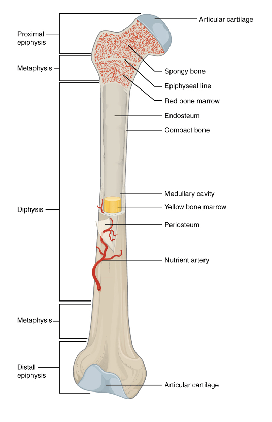
Question 71:
**Answer: C) Appositional growth**
**Explanation:**
Osteoblasts contribute to bone growth through appositional growth. Appositional growth involves the increase in bone thickness or diameter, achieved by the addition of new bone tissue on the bone’s surface by osteoblasts (from the periosteum & then bone is added inwards).
Interstitial growth refers to the increase in length of bones, which typically occurs at the epiphyseal plates during development. This process involves chondrocytes (cartilage-forming cells) stacking up on top of one another to increase the length of the bone.
Endochondral ossification is a process of bone formation in which cartilage is replaced by bone; it is not a direct mechanism of osteoblast growth but rather a developmental process involving chondrocytes.
Intramembranous ossification is a process involving the direct formation of bone within a connective tissue membrane from mesenchymal cells, contributing to the formation of flat bones like those of the skull. It is a bone formation process rather than a specific growth mechanism of osteoblasts.
Calcification refers to the deposition of calcium salts within the tissue, which hardens the bone matrix. It is a part of bone formation but not a direct growth mechanism of osteoblasts.

Question 72:
**Answer: E) Diarthrosis**
**Explanation:**
Diarthroses aka synovial joints, allow for a wide range of motion in various directions, such as the shoulder, hip, and knee joints.
Gomphosis is a type of fibrous joint where a peg fits into a socket, such as the joint between a tooth and its socket in the jaw, which is immovable (synarthrosis).
Syndesmosis is a type of fibrous joint where bones are connected by a ligament or an interosseous membrane, allowing for slight movement, making it an amphiarthrosis rather than a freely moveable joint.
Synarthrosis refers to an immovable joint, such as the sutures in the skull (fibrous joints).
Amphiarthrosis describes a joint that allows for limited movement, such as the intervertebral discs in the spine or the pubic symphysis (secondary cartilaginous joints).
Ways to remember the following:
- Synarthrosis: Think sin. When someone commits a major sin, they might be punished & as a result can never move again = no movement
- Diarthrosis: Think diarrhoea, when you have diarrhoea it goes everywhere = full range of movement
- Amphiarthrosis: Think amphibolic reaction which is a reaction that can go both ways (i.e. a catabolic & anabolic reaction) = not too much & not no movement so there is a small range of movements here.

Question 73:
**Answer: D) 1st rib & sternum**
**Explanation:**
Primary cartilaginous joints, also known as synchondroses, are characterised by hyaline cartilage directly connecting the bones, allowing for no movement, and typically found in developing bones (synarthrosis). Other examples of primary cartilaginous joints are the joint between the sphenoid & occipital bone in the skull & the joint between the diaphysis & epiphysis.
Fibrous Joints (synarthrosis):
- Sutures: These are joints between the bones of the skull, connected by dense fibrous connective tissue. They are typically immovable.
- Syndesmosis: This type involves a membrane or ligament connecting two bones, such as the tibia and fibula or the radius and ulna. It allows for slight movement.
- Gomphosis: This is a peg-and-socket joint found between the teeth and their sockets in the jaw. It is also immovable.
Secondary Cartilaginous Joints (Symphyses aka amphiarthrosis):
These joints involve a layer of fibrocartilage between two layers of hyaline cartilage. They are slightly movable. Examples include the pubic symphysis, intervertebral discs, and the manubriosternal joint. The fibrocartilage provides cushioning and allows for slight movement, which is more than that found in primary cartilaginous joints.

Question 74:
**Answer: C) Bursa & fat pads**
**Explanation:**
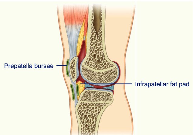
Intra-articular structures are those located within the joint capsule, directly interacting with the joint space between bones. Bursa & fat pads are not located within the joint capsule. Bursae are fluid-filled sacs that reduce friction between moving structures, and fat pads provide cushioning around the joint. These structures are located adjacent to the joint but are not within the intra-articular space.
Menisci are crescent-shaped cartilage structures found within certain joints, such as the knee. They are intra-articular because they are located within the joint space, where they aid in load distribution and joint stability.
Intra-articular discs are fibrocartilaginous structures found within the joint capsule, such as those in the temporomandibular joint. They are considered intra-articular as they reside within the joint space and help with load distribution and joint congruency.
Intra-articular ligaments are located within the joint capsule and contribute to the stability and support of the joint. An example is the anterior cruciate ligament (ACL) in the knee.
Labra (plural of labrum) are cartilaginous structures that deepen the sockets of ball-and-socket joints, such as the shoulder and hip. They are intra-articular as they are located within the joint capsule, enhancing joint stability.
Question 75:
**Answer: A) Circumduction**
**Explanation:**
Circumduction is a circular movement that involves a combination of flexion, extension, abduction, and adduction. This movement is typically seen in ball-and-socket joints, such as the shoulder or hip, and is not possible at the foot, where movements are more restricted to the planes of flexion and extension or inversion and eversion.
Dorsiflexion refers to the movement where the foot is brought closer to the shin/pointing upwards towards the sky, effectively decreasing the angle between the dorsum (top) of the foot and the leg.
Plantarflexion is the opposite of dorsiflexion, involving the movement where the foot is pointed downward towards the floor, increasing the angle between the dorsum of the foot and the leg.
Inversion is the movement where the sole of the foot turns inward, towards the midline of the body.
Eversion is the movement where the sole of the foot turns outward, away from the midline of the body.
Question 76:
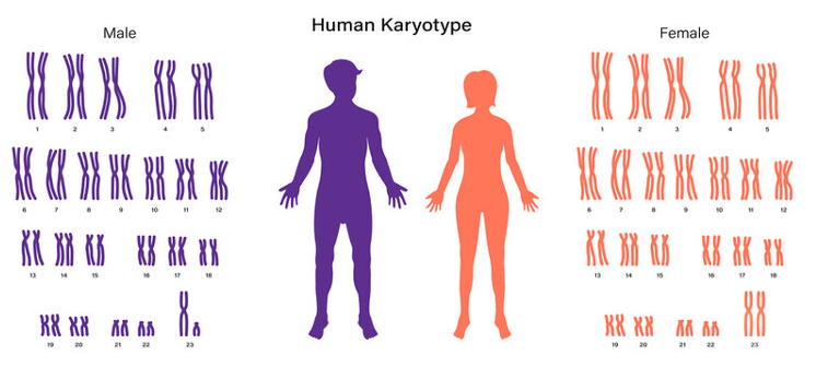
**Answer: D) Karyotype**
**Explanation:**
The complete set of ordered chromosomes refers to the organised arrangement of chromosomes in a cell, typically displayed in a karyogram or karyotype, which provides a comprehensive view of an organism’s chromosomal makeup, typically arranged by size, shape, and number, providing a systematic representation of the chromosomal complement.
Genotype refers to the genetic constitution of an individual, specifically the alleles present at a particular gene or set of genes.
Phenotype is the observable characteristics or traits of an individual, resulting from the interaction between the genotype and the environment.
Proteome is the entire set of proteins expressed by a genome, cell, tissue, or organism at a certain time.
Prototype is a term used to describe an original model or preliminary version of a product or concept, not related to chromosomes.
Question 77:
**Answer: D) Nucleosome**
**Explanation:**
Nucleosome is the correct term for the complete structure that includes DNA wrapped around a core of 8 histone proteins. This is the fundamental unit of chromatin.
An octamer refers specifically to the core of 8 histone proteins around which DNA wraps, but it does not include the DNA itself, making it incomplete as a term for the entire complex.
Chromatin fibre describes a higher-order structure that consists of multiple nucleosomes arranged along the DNA, but it is not the fundamental unit formed by the DNA-histone interaction.
Histone core refers to the histone proteins around which DNA wraps, but it does not encompass the DNA wrapped around these proteins.
Scaffold refers to the protein framework that helps organize and support the higher-order structure of chromatin but does not specifically describe the DNA-histone complex.

Question 78:
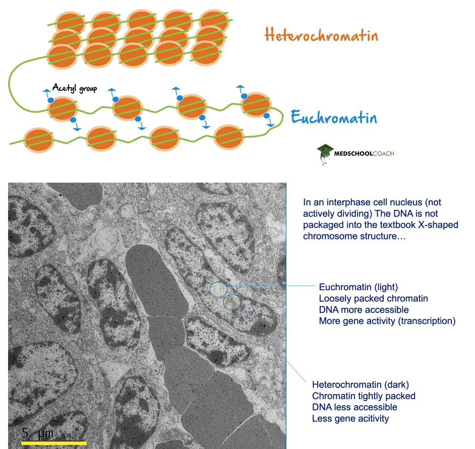
**Answer: A) Euchromatin**
**Explanation:**
Euchromatin refers to the loosely packed form of chromatin that allows for easier access to DNA, making it more available for transcription. This results in higher levels of gene expression.
Heterochromatin, on the other hand, is the tightly packed form of chromatin that is less accessible and typically associated with lower levels of transcription.
Solenoid refers to a higher-order structure of chromatin where nucleosomes are organised into a coiled structure. It does not specifically describe the accessibility of DNA for transcription.
Scaffold chromatin refers to the protein framework that organises the chromatin into its higher-order structures but does not directly describe the accessibility of DNA for transcription.
Chromatin loop is a term that describes the looped structure of chromatin domains within the nucleus, which helps in organising and regulating gene expression but does not directly address the level of DNA accessibility for transcription.
Question 79:
**Answer: E) Sickle cell anaemia**
**Explanation:**
Sickle cell anaemia is a genetic disorder caused by an autosomal recessive phenotype. This means that two copies of the mutated gene (one from each parent) are required for the disease to manifest. The condition is characterised by the production of abnormal haemoglobin, leading to distorted red blood cells that can cause various complications.
Huntington’s disease is an autosomal dominant disorder where only one copy of the mutated gene is sufficient to cause the disease. It leads to neurodegenerative symptoms that usually appear in mid-adulthood.
Retinoblastoma is an autosomal dominant cancer of the retina. It requires only one copy of the mutated gene to increase the risk of developing the disease, although it can sometimes be inherited in a recessive manner.
Polycystic kidney disease is typically autosomal dominant. It results in the formation of cysts in the kidneys and requires only one copy of the mutated gene for the disease to develop.
Marfan Syndrome is another autosomal dominant disorder that affects connective tissue, resulting in features like tall stature and cardiovascular issues.
Question 80:
**Answer: B) Autosomal dominant**
**Explanation:**
In autosomal dominant conditions, the disease often does NOT skip generations, i.e. in every single generation, you should find an affected individual. If someone’s parent is affected, the person has a 50% chance of inheriting the dominant allele.
How to spot the remaining inheritance patterns?
Autosomal recessive = the disease often skips generations i.e. one generation will have the disease whilst the next will not (accept if both parents have the disease therefore all children should be affected). If someone’s parent is affected, there is a 25% chance that the person will inherit both recessive alleles.
Mitochondrial = if mother is affected, all children will have the disease since mitochondria is inherited maternally not paternally. If the father has a mitochondrial disease, he can NOT pass it on to his offspring. If someone’s mother is affected, there is 100% chance that they will be affected too.
X-linked recessive =
- More Males Affected: Males are more frequently affected; they inherit the condition from their mothers.
- Carrier Mothers: Affected males usually have carrier mothers.
- Can Skip Generations: The trait may not appear in every generation due to female carriers.
- No Father-to-Son Transmission: Affected fathers do not pass the trait to their sons.
X-linked dominant =
- More Affected Females: Females are more commonly affected; both males and females with the dominant allele show the condition.
- Affected Fathers Pass to Daughters: Affected fathers pass the trait to all their daughters.
- No Carriers: If a female has the dominant allele, she will express the condition.
- No Skipping Generations: The condition usually appears in every generation.
Question 81:
**Answer: D) Adenine & thymine**
**Explanation:**
In DNA, complementary base pairing follows specific rules where each base pairs with a corresponding partner:
- Adenine (A) pairs with Thymine (T) through two hydrogen bonds.
- Cytosine (C) pairs with Guanine (G) through three hydrogen bonds.

Question 82:
**Answer: E) Ligase**
**Explanation:**
Okazaki fragments are short segments of DNA synthesised on the lagging strand during DNA replication. These fragments are initially created by DNA polymerase but must be joined together to form a continuous strand. Ligase is the enzyme responsible for joining Okazaki fragments together by sealing the nicks in the DNA backbone, creating a continuous strand.
DNA helicase unwinds the DNA double helix, separating the strands for replication, but does not join Okazaki fragments.
Taq polymerase is a heat-stable enzyme used in PCR to synthesise & elongate DNA by adding nucleotides to the existing template strand.
Telomerase extends the telomeres of chromosomes to prevent loss of genetic material during replication.
Primase synthesises short RNA primers needed for DNA polymerase to begin synthesis.
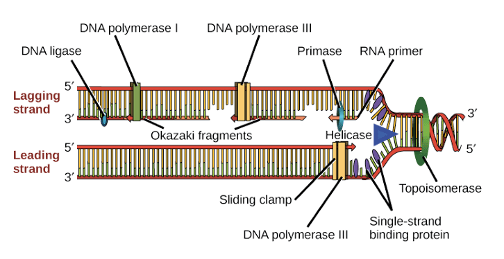
Question 83:
**Answer: C) rRNA**
**Explanation:**
Ribosomes are complex molecular machines responsible for protein synthesis in cells (during translation). Ribosomes are synthesised in the nucleolus of the cell, where rRNA and ribosomal proteins are combined to form ribosomal subunits.
DNA provides the genetic instructions for the synthesis of mRNA (transcription).
RNA serves as a broader category that includes several types of RNA (such as rRNA, tRNA & mRNA), but it is not specific enough.
mRNA (messenger RNA) carries genetic information from DNA to the ribosome but is not involved in ribosome synthesis.
tRNA (transfer RNA) aids in translating mRNA into proteins/polypeptide chains (during translation) by carrying & bringing a specific amino acid to the ribosome.
Question 84:
**Answer: A) Kinesin**
**Explanation:**
In cells, the transport of molecules and organelles along the cytoskeleton involves motor proteins that move along specific tracks. Antegrade transport, which is the forward movement of materials toward the cell’s periphery, relies on specific structures. Kinesin is the motor protein responsible for antegrade (forward) transport along microtubules, carrying cargo from the cell body toward the cell periphery.
Dynein, on the other hand, is involved in retrograde (backward) transport, moving materials from the cell periphery back to the cell body. Retro = past so dyneins are involved in the transport of cargo backwards. ‘D’ rhymes with ‘B’ so dynein = backwards.
Microtubules are the structural tracks along which motor proteins like kinesin and dynein move.
Tubulin is the protein subunit that makes up microtubules but is not directly responsible for transport itself.
Myosin is a motor protein that interacts with actin filaments, not microtubules, and is involved in processes such as muscle contraction and cell motility rather than antegrade transport along microtubules.
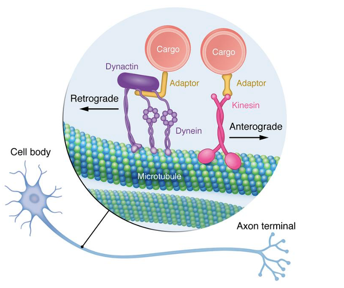
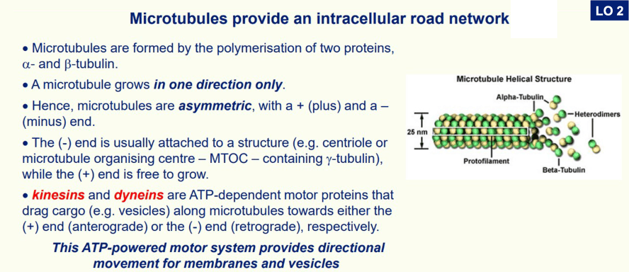
Question 85:
**Answer: B) Actin cytoskeleton**
**Explanation:**
To move the entire cell, various cytoskeletal structures and their associated proteins play critical roles. The actin cytoskeleton, consisting of actin filaments (including both F-actin and G-actin), provides the structural framework necessary for cell motility and shape changes. This network is essential for processes like amoeboid movement, muscle contraction, and cell crawling.
Microtubules are primarily involved in intracellular transport and cell division rather than in moving the entire cell.
Actin filaments, or microfilaments, are part of the actin cytoskeleton, which is crucial for cell movement.
F-actin and G-actin are forms of actin: F-actin is the filamentous form, while G-actin is the globular monomeric form. While both forms are involved in actin polymerisation and dynamics, the term “actin cytoskeleton” encompasses the entire network of actin filaments that are critical for cell movement.

Question 86:
**Answer: B) Secondary oocyte**
**Explanation:**
Oogenesis is the process of egg (ovum) development in females, involving two major stages of meiosis. Following meiosis I, the primary oocyte, which has undergone DNA replication and entered prophase I, completes meiosis I to produce two cells.
The two structures formed are:
- Secondary oocyte: This is the larger cell that receives most of the cytoplasm and is arrested in metaphase II of meiosis until fertilisation.
- Polar body: This smaller cell, which receives minimal cytoplasm and is typically non-functional, is a by-product of meiosis I and is eventually discarded.
The primary oocyte is the cell that initiates meiosis but does not complete meiosis I until later in life. It produced following DNA replication in mitosis.
Spermatozoa are the mature male gametes, and the primary spermatocyte is a male cell that undergoes meiosis to form sperm cells.
Oogenesis is the process of egg (ovum) development in females and involves several key stages:
- Fetal Development: Primordial germ cells develop into primary oocytes. These oocytes begin meiosis but pause in prophase I until puberty.
- Puberty: Each menstrual cycle, a primary oocyte resumes meiosis. It completes meiosis I, producing a secondary oocyte and a smaller polar body.
- Secondary Oocyte: The secondary oocyte progresses to metaphase II of meiosis but stops there until fertilisation.
- Fertilisation: If fertilisation occurs, the secondary oocyte completes meiosis II, producing a final ovum and a second polar body.
- Polar Bodies: These are small, non-functional cells produced during meiosis, which eventually degenerate.
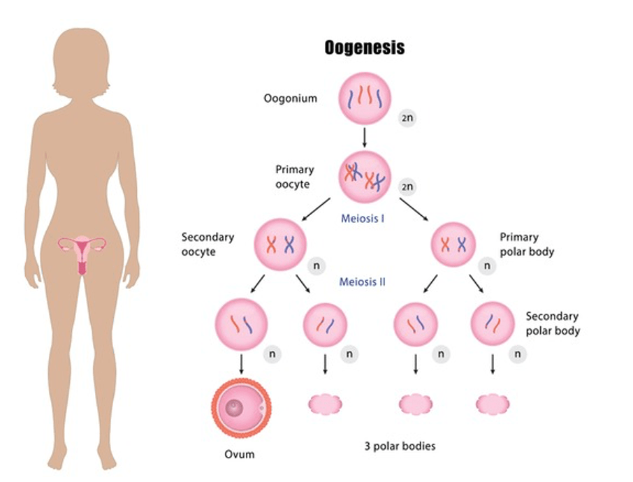
Question 87:
**Answer: A) Spermatozoa**
**Explanation:**
Spermatogenesis is the process of sperm development in males, involving several stages of maturation:
- Spermatogonia are the initial germ cells that divide by mitosis to produce primary spermatocytes.
- Primary spermatocytes undergo meiosis I to form secondary spermatocytes.
- Secondary spermatocytes then complete meiosis II to produce spermatids.
- Spermatids are the intermediate cells that undergo a transformation process called spermiogenesis to become mature sperm aka spermatozoa.

Question 88:
**Answer: D) Polyploidy**
**Explanation:**
Polyploidy is the condition where cells have more than 2 complete sets of chromosomes (no longer a diploid chromosomes in autosomal cell & haploid chromosomes in a sex cell). This means that cells contain more than 2 pairs of homologous chromosomes, which can occur in plants and some animal species g. 3N, 4N, 5N etc. & this is lethal in humans.
Aneuploidy refers to the presence of an abnormal number of chromosomes in a cell e.g., 45 or 47 chromosomes rather than 46.
Penetrance is the proportion of individuals with a particular genotype that actually displays the associated phenotype.
Incomplete penetrance occurs when not all individuals with a genotype express the expected phenotype.
Lyonization, also known as X-inactivation, is the process by which one of the two X chromosomes in female mammals is randomly inactivated during early embryonic development.
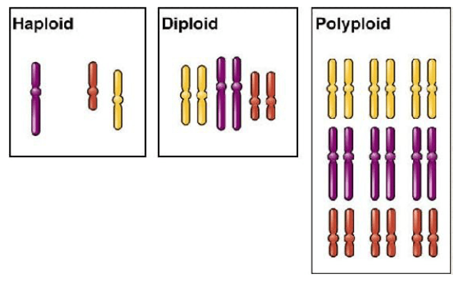
Question 89:
**Answer: B) Prophase I until the start of the menstrual cycle**
**Explanation:**
1-2 months before the birth of a female baby, most of her 7 million oogonia die, and the remaining surviving oogonia enter meiosis I and become primary oocytes. Primary oocytes begin meiosis during fetal development but then arrest in prophase I (stop dividing). They remain arrested in this stage until puberty. With the onset of puberty and the start of the menstrual cycle, one primary oocyte resumes meiosis during each cycle & becomes a secondary oocyte. However, the secondary oocyte arrests at metaphase II until it is fertilised.
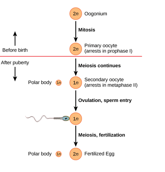
Question 90:
**Answer: C) Stem cells differentiate into absorptive (villous) cells**
**Explanation:**
The Notch signalling pathway is crucial for cell fate determination in various tissues, including the intestine. In the colonic crypt, Notch signalling plays a key role in determining whether stem cells will differentiate into absorptive (villous) cells in the intestines or secretory cells (like goblet, enteroendocrine, and Paneth cells in the colonic crypt).
When the Notch ligand is activated or “ON,” it promotes the differentiation of stem cells into absorptive cells, which are the villous cells responsible for nutrient absorption. This activation inhibits the differentiation into secretory cell lineages. When Notch ligand is inactivated or turned “OFF”, stem cells differentiate into secretory cells (colonic cells).


Question 91:
**Answer: E) Keratinocytes**
**Explanation:**
Keratinocytes are the primary cell type found in the epidermis of the skin. They produce keratin, a protein that helps protect the skin from damage, dehydration, and pathogens. These are self-regenerating stratified squamous cells.
Hepatocytes are the main cell type found in the liver, responsible for various metabolic processes.
Langerhans cells are a type of dendritic cell involved in the immune response and are found in the skin’s epidermis but are not the primary cell type.
Chondrocytes are the cells found in cartilage.
Enterocytes are the absorptive cells in the lining of the intestines.
Question 92:
**Answer: A) Merkel cell**
**Explanation:**
Merkel cells are found in the skin and function as mechanoreceptors, which means they are involved in the sensation of touch. These cells are located in the basal layer of the epidermis and are associated with nerve endings to help detect light touch and pressure.
Mast cells are involved in the immune response and are found in connective tissues throughout the body, including the skin.
Dendritic cells, such as Langerhans cells, are also involved in the immune response and are present in the skin but do not function as mechanoreceptors.
Melanocytes are responsible for producing melanin, the pigment that gives skin its colour.
Basophils are a type of white blood cell involved in the immune response (by releasing histamine).
Question 93:
**Answer: C) Monomer**
**Explanation:**
Cyclins and cyclin-dependent kinases (CDKs) are crucial regulatory proteins involved in regulating the cell cycle (via checkpoints). Each cyclin and CDK is individually a monomer, meaning they exist as single, unbound molecules. When cyclins bind to CDKs, they form an active complex that can then phosphorylate target proteins to drive cell cycle progression.
Multimer refers to a complex of more than two molecules. Polymer refers to a large molecule made up of repeating subunits.
Dimer refers to a complex of two molecules bound together, which describes the functional state when a cyclin binds to a CDK, but not their individual forms.
Oligomer refers to a complex of a few molecules, between 10-20, which is also not the primary form of cyclins and CDKs.
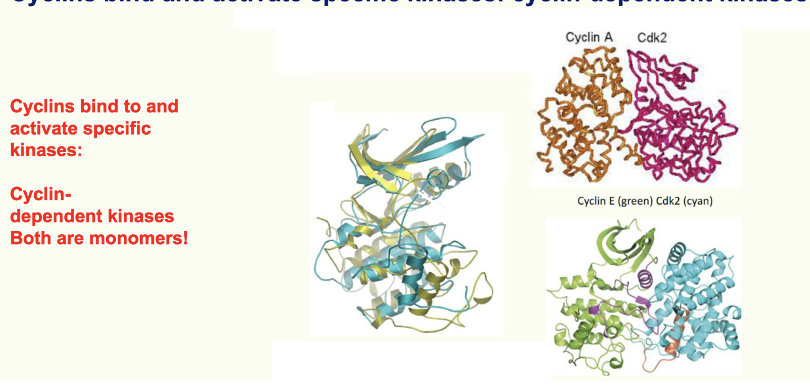
Question 94:
**Answer: D) Tetramer**
**Explanation:**
p53 is a tumour suppressor protein that plays a crucial role in preventing cancer by regulating the cell cycle and inducing apoptosis in response to DNA damage. The functional form of p53 is a tetramer, meaning it consists of four subunits.
Monomer refers to a single molecule that is not bound to other molecules. Dimer refers to a complex of two molecules. Trimer refers to a complex of three molecules. Tetramer refers to a complex of four molecules, which is the correct form for functional p53. Pentamer refers to a complex of five molecules.
Question 95:
**Answer: C) Amphibolic reaction**
**Explanation:**
The reaction in which products can be both made (synthesized) and broken down (decomposed) is called an amphibolic reaction. Amphibolic pathways are those that involve both catabolic and anabolic processes, allowing the same metabolic pathways to function in both directions depending on cellular needs and conditions.
A catabolic reaction refers to reactions that break down complex molecules into simpler ones, releasing energy.
An anabolic reaction refers to reactions that build complex molecules from simpler ones, requiring energy.
An aqueous reaction refers to reactions that occur in a water-based environment.
An allosteric reaction refers to the regulation of enzyme activity through binding at a site other than the active site.
Question 96:
**Answer: B) Beta oxidation**
**Explanation:**
Beta oxidation is the process in which fatty acids are broken down in the mitochondria to generate AcetylCoA, which can then enter the citric acid cycle for further energy production.
Gluconeogenesis is the process of synthesising glucose from non-carbohydrate sources such as glycerol & amino acids.
Substrate level phosphorylation is a method of generating ATP during metabolic reactions (from ADP + Pi).
Lipolysis refers to the breakdown of triglycerides into glycerol and free fatty acids.
Lipogenesis is the process of synthesising fatty acids from acetyl-CoA and other substrates.
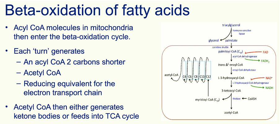
Question 97:
**Answer: A) Essential amino acids cannot be synthesised by the body (need to be obtained by diet) whereas non-essential amino acids can be synthesised by the body.**
**Explanation:**
Essential amino acids are crucial for protein synthesis and various metabolic processes, and their absence in the diet can lead to deficiencies. Non-essential amino acids, although also important, can be produced internally and thus do not need to be obtained directly from the diet.

Question 98:
**Answer: E) cAMP**
**Explanation:**
In the Gs signalling pathway of G-protein coupled receptors, adenylate cyclase is activated by the Gs protein, leading to the conversion of ATP into cyclic AMP (cAMP), which acts as a second messenger. cAMP then activates protein kinase A (PKA) to elicit various cellular responses.
Phospholipase C is an enzyme involved in a different signalling pathway, specifically the Gq pathway.
Calcium ions are second messengers involved in the Gq signalling pathway which trigger the release of calmodulin.
Adenylate cyclase is an enzyme that produces cAMP but is not itself a second messenger.

Question 99:
**Answer: B) Ependymal cells**
**Explanation:**
Ependymal cells are a type of glial cell that line the ventricles of the brain and the central canal of the spinal cord. They are involved in the production and circulation of cerebrospinal fluid (CSF).
Astrocytes support neurons and maintain the blood-brain barrier.
Microglial cells act as the brain’s immune cells.
Oligodendrocytes myelinate to axons in the central nervous system.
Neurons are the primary signalling cells of the nervous system and are not classified as glial cells.
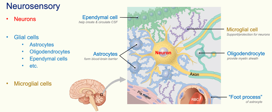
Question 100:
**Answer: A) 1**
**Explanation:**
There is typically one spinal nerve in the coccyx region, known as the coccygeal nerve. This nerve emerges from the coccygeal segment of the spinal cord and is responsible for sensory and motor functions in the coccygeal area.
There are 3-4 fused coccygeal bones but only 1 nerve.
There are 5 lumbar & 5 sacral nerves.
There are 8 cervical nerves & 12 thoracic nerves.
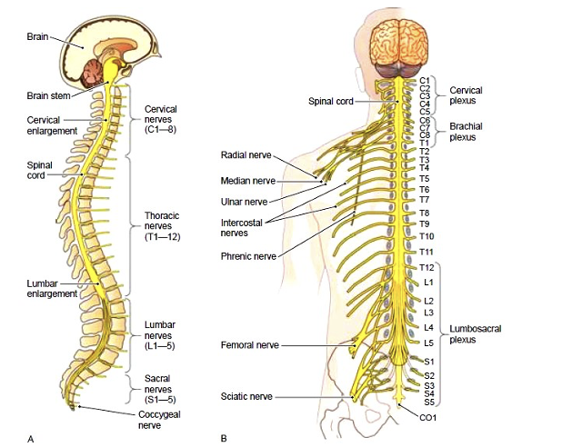
Question 101:
**Answer: D) Dorsal root**
**Explanation:**
Afferent neurons, which carry sensory information from the peripheral nervous system to the central nervous system, enter the spinal cord via the dorsal root. The dorsal root contains the axons of sensory neurons and is responsible for transmitting sensory signals to the central nervous system.
The ventral root carries efferent motor signals from the central nervous system to the muscles and glands.
The ventral ramus and dorsal ramus are branches of a spinal nerve that emerge from the spinal cord:
- Dorsal ramus: This branch is closer to the spinal cord and is smaller. It immediately branches off from the spinal nerve and provides motor & sensory innervation to the back of the body, including the skin and muscles along the spine.
- Ventral ramus: This branch is larger and also emerges directly from the spinal nerve but provides motor & sensory innervation to the front and sides of the body. It branches further to supply the limbs and the anterior parts of the trunk.
The dorsal root ganglion contains the cell bodies of sensory neurons, but the actual axons of these neurons enter the central nervous system through the dorsal root.
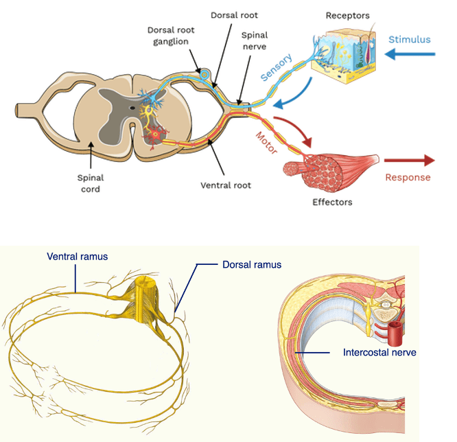
Question 102:
**Answer: D) Trigeminal **
**Explanation:**
The trigeminal nerve, which is the 5th cranial nerve and is responsible for sensation in the face and provides motor innervation to the muscles of mastication adding in chewing.
The optic nerve is the 2nd cranial nerve and is responsible for vision.
The glossopharyngeal nerve is the 9th cranial nerve, involved in taste, salivation, and throat sensation.
The accessory nerve is the 11th cranial nerve, responsible for motor functions in the neck and shoulders.
The vestibulocochlear nerve is the 8th cranial nerve, which handles hearing and balance.

Question 103:
**Answer: A) Fascial nerve**
**Explanation:**
The facial nerve provides both sensory and motor innervation. It controls the muscles of facial expression, provides taste sensation to the anterior two-thirds of the tongue & innervates the submandibular & sublingual salivary glands.
The hypoglossal nerve (CN XII) is purely motor, innervating the tongue muscles (allows tongue to move).
The trochlear nerve (CN IV) and the abducens nerve (CN VI) are purely motor nerves, controlling eye movements (superior oblique muscle & lateral rectus muscle respectively).
The olfactory nerve (CN I) is purely sensory and is involved in the sense of smell.
Remember the mnemonic: Some Say Marry Money But My Brother Says Big Brains Matter More.
S – Sensory. M – Motor. B – Both

Question 104:
**Answer: B) C5-T1**
**Explanation:**
The brachial plexus is formed from the spinal roots of the cervical nerves C5 through C8 and the first thoracic nerve (T1). These roots combine to form the trunks, divisions, branches and cords of the brachial plexus, which innervate the shoulder, arm, and hand.
C4-T1 is incorrect because it includes C4, which is not part of the brachial plexus. The brachial plexus starts at C5.
S2-S4 refers to the sacral plexus, which innervates the pelvic region and lower limbs, not the brachial plexus.
L1-L5 refers to the lumbar plexus, which serves the lower abdomen, groin, and parts of the lower limbs, not the brachial plexus.
C1-C4, refers to the cervical plexus, which innervates the neck and parts of the diaphragm, not the brachial plexus.
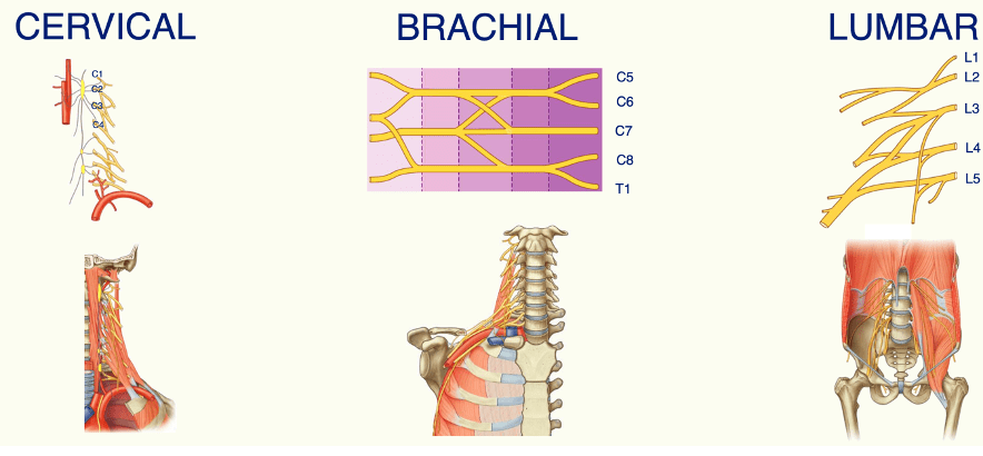
Question 105:
**Answer: E) The sympathetic nervous system has a short preganglionic and long postganglionic neuron. Whereas the parasympathetic nervous system has a long preganglionic and a short postganglionic neuron.**
**Explanation:**
In the sympathetic nervous system, the preganglionic neurons are short (close to the CNS) and synapse with postganglionic neurons that are long, which is why sympathetic responses are typically more widespread. In contrast, in the parasympathetic nervous system, the preganglionic neurons are long and synapse with short postganglionic neurons (close to visceral organs), leading to more localized and specific responses.
Sympathetic neurons exit the spinal cord at thoracic & lumbar regions, T1-L2. Whereas parasympathetic neurons exit the CNS at the cranium (brain) & the sacral region of the spinal cord.
Options A and B are incorrect as both systems have two neurons involved in a single pathway.
Option C is correct but does not capture the key difference in neuron length.
Option D incorrectly describes the relative lengths of the neurons in the two systems.
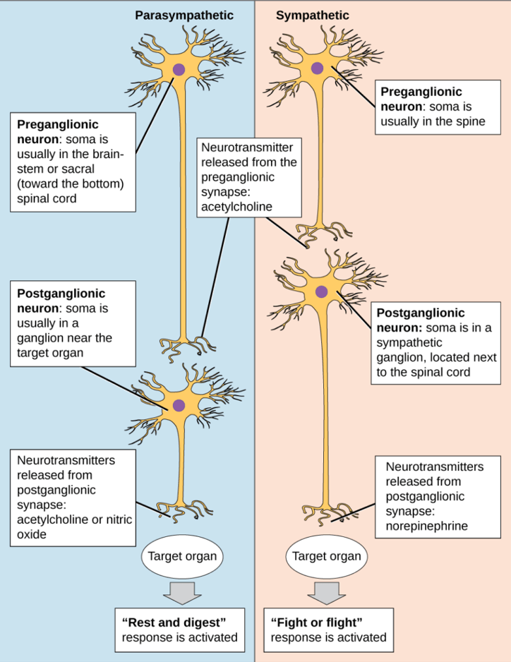
Question 106:
**Answer: A) Ultrasound scans**
**Explanation:**
Ultrasound is considered the safest imaging technique for paediatric patients because it does not use ionising radiation, making it preferable for frequent imaging in children. It is also mobile so can be transported to the patient if they are unable to move.
MRI scans are also safe and do not use ionising radiation, but they are less commonly used due to higher costs and longer scan times which require the patient to lay still for an extended period of time.
CT scans and X-rays involve ionising radiation, which carries a higher risk, especially with frequent exposure.
Fluoroscopy also uses ionising radiation and is less commonly used in paediatrics due to its potential risks.
Question 107:
**Answer: A) Collagen type I**
**Explanation:**
Tendons & ligaments are primarily composed of collagen type I, which provides strength and structural support. This type of collagen is the most abundant in the body and is crucial for the tensile strength of tendons.
Collagen type II is mainly found in cartilage, collagen type III is found in skin, blood vessels & following bone damage.
Collagen type IV is a major component of the basement membrane, and collagen type V is found in small amounts in various tissues, including tendons, but is not the primary type.
Question 108:
**Answer: C) T-tubules**
**Explanation:**
T-tubules (transverse tubules) are extensions of the muscle cell membrane (sarcolemma) that penetrate into the interior of the muscle fibre. They conduct action potentials from the surface of the muscle cell deep into the myocyte, ensuring that the electrical signal reaches all parts of the muscle fibre.
The sarcoplasmic reticulum stores calcium ions and releases them in response to action potentials, but it does not directly carry action potentials.
Gap junctions allow for electrical communication between adjacent cells but are not involved in carrying action potentials into the myocyte.
Calcium ions are involved in muscle contraction but do not convey action potentials themselves.
Desmosomes are involved in cell adhesion, not in action potential conduction.
Question 109:
**Answer: E) High concentration of glycogen**
**Explanation:**
Type I slow-twitch fibers are designed for endurance and prolonged activity. They are red in colour due to a high concentration of myoglobin, have an abundant number of mitochondria, and generate ATP primarily through oxidative phosphorylation, which allows for sustained energy release and endurance. Type I fibers generally have a lower glycogen content because they primarily rely on aerobic metabolism.
A high concentration of glycogen is characteristic of type II fast-twitch fibers, which are designed for quick, powerful bursts of activity and rely more on glycolytic pathways for energy.
Question 110:
**Answer: B) 7.35-7.45**
**Explanation:**
This slight alkalinity is crucial for the proper functioning of various physiological processes in the body.
Question 111:
**Answer: C) Yolk sac**
**Explanation:**
Yolk Sac (Early Embryonic Stage): During the first few weeks of gestation, the yolk sac is the initial site of blood cell production. It provides primitive haematopoiesis, producing early blood cells and blood vessels between 3 to 8 weeks of gestation.
Liver (Mid-Gestation): After the yolk sac, the liver becomes the main site of haematopoiesis and is responsible for producing a large number of red blood cells, white blood cells, and platelets. From about 6 weeks to birth, the liver is the predominant site.
Spleen (Mid to Late Gestation): Alongside the liver, the spleen contributes to haematopoiesis, particularly in the production of lymphocytes and other immune cells between 10 to 28 weeks of gestation.
Bone Marrow (Late Fetal Development): Haematopoiesis gradually shifts to the bone marrow as the fetus develops, becoming the primary site by birth. The red bone marrow takes over the production of all blood cell types. Starts around 20 weeks and becomes the main site by birth.

Question 112:
**Answer: D) Neutrophil**
**Explanation:**
Neutrophils are a type of white blood cell and the most abundant type of granulocytes in the blood. They are characterised by a multi-lobed nucleus, typically with three to five lobes connected by thin strands of chromatin. This unique structure allows them to move flexibly through tissues. Neutrophils are specially designed to destroy bacteria and fungi through processes such as phagocytosis, degranulation, and the release of antimicrobial enzymes.
A lymphocyte is a type of white blood cell involved in the adaptive immune response. They have a large, round nucleus and are responsible for antibody production and immune regulation.
Basophils are another type of granulocyte with a bilobed nucleus. They play a role in allergic reactions and inflammation by releasing histamine and other chemicals.
Macrophages are large phagocytic cells that originate from monocytes. They have a single, large, kidney-shaped nucleus and are involved in engulfing pathogens and debris. While they are capable of destroying bacteria, they do not have a multi-lobed nucleus like neutrophils.
A phagocyte is a general term for any cell that engulfs and digests foreign particles, bacteria, and dead or dying cells. While neutrophils are a type of phagocyte, this term is too broad and does not specifically refer to the multi-lobed nucleus characteristic.
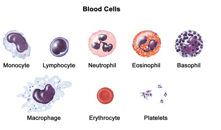
Question 113:
**Answer: E) Size of cell**
**Explanation:**
Forward scatter (FSC) in flow cytometry is primarily used to measure the size of a cell. As cells pass through the laser beam in a flow cytometer, the amount of light scattered in the forward direction is proportional to the cell’s size. Larger cells scatter more light forward than smaller cells, allowing researchers to distinguish between different cell populations based on size.
Cell complexity is measured by side scatter (SSC), not forward scatter. Side scatter provides information about the granularity or internal complexity of a cell, such as the presence of granules and the complexity of the internal structure.
Volume of cytoplasm is not directly measured by forward scatter. While cell size can indirectly suggest cytoplasmic volume, forward scatter primarily detects overall size rather than specific cellular components.
The range of surface proteins is detected using specific fluorescently labelled antibodies in flow cytometry. These proteins are not directly measured by forward scatter.
The total number of cells present is determined by counting events as they pass through the flow cytometer.
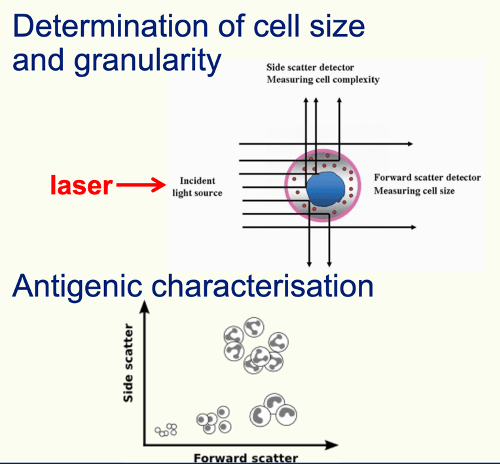
Question 114:
**Answer: A) CD45**
**Explanation:**
CD45, also known as leukocyte common antigen (LCA), is a protein tyrosine phosphatase expressed on the surface of all leukocytes, including lymphocytes, monocytes, eosinophils, basophils, and neutrophils. It plays a critical role in leukocyte activation and signal transduction. This marker is widely used in flow cytometry to identify and analyse leukocyte populations.
CD34 is a marker found on hematopoietic stem cells and early progenitor cells, as well as endothelial cells, but it is not present on all leukocytes. It is often used to identify stem cells in bone marrow transplants.
CD1 is a marker associated with antigen-presenting cells such as dendritic cells and certain subsets of T cells, but not all leukocytes.
CD3 is a marker specific to T lymphocytes, as it is part of the T cell receptor (TCR) complex and is not found on other types of leukocytes like B cells, monocytes, or granulocytes.
CD8 is a marker found on cytotoxic T cells, a subset of T lymphocytes, and not on all leukocytes. CD8 is involved in the recognition of antigens presented by MHC class I molecules.

Question 115:
**Answer: C) Natural killer cell**
**Explanation:**
Natural killer (NK) cells are classified as lymphocytes because of their lineage but function within the innate immune system. They are involved in the body’s early defence against infected or cancerous cells by identifying and destroying them without the need for prior exposure.
T lymphocytes (Option A) and B lymphocytes (Option B) are part of the adaptive immune system. T cells are involved in cell-mediated immunity and B cells in humoral immunity (antibody production), both requiring antigen presentation and prior exposure to specific pathogens.
Dendritic cell are antigen-presenting cells that help bridge the innate and adaptive immune systems but are not classified as lymphocytes.
Megakaryocytes are not leukocytes; they are large bone marrow cells responsible for producing platelets involved in blood clotting.
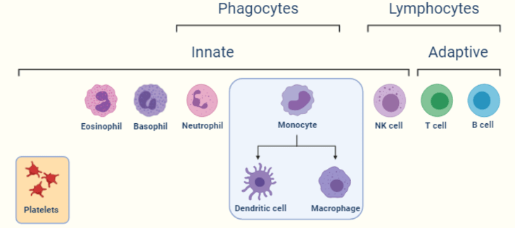
Question 116:
**Answer: B) AID enzyme**
**Explanation:**
The Activation-Induced Cytidine Deaminase (AID) enzyme is essential for antibody class-switch recombination. AID works by deaminating cytosine residues in DNA, leading to mutations and double-stranded DNA breaks. These changes are crucial for the recombination process that allows B cells to switch the class of antibody they produce (such as from IgM to IgG, IgA, or IgE) while maintaining specificity for the same antigen.
RAG enzyme: The Recombination Activating Genes (RAG-1 and RAG-2) are involved in V(D)J recombination, the process that creates the diverse repertoire of antigen receptors on B and T cells. RAG enzymes are crucial for generating diversity in antigen receptors but are not involved in the class-switch recombination process.
VDJ recombinase: This term generally refers to the RAG enzymes, which are involved in V(D)J recombination. While they are important for the initial assembly of antigen receptors, they do not play a role in class-switch recombination.
Terminal deoxynucleotidyl transferase (TdT): TdT adds nucleotides to the ends of DNA molecules, particularly during the rearrangement of antigen receptor genes. While TdT is important for diversity in antigen receptors, it is not specifically involved in antibody class-switch recombination.
DNA ligase is an enzyme that joins DNA strands by forming phosphodiester bonds between adjacent nucleotides. It plays a role in various DNA repair and recombination processes, including some final steps of recombination, but is not specifically required for class-switch recombination.
Question 117:
**Answer: D) Stratum spinosum**
**Explanation:** Deep to superficial-
- Stratum basale – the deepest layer where new skin cells are generated.
- Stratum spinosum – the second layer where cells have a spiny appearance due to desmosomes.
- Stratum granulosum – the third layer where cells start to undergo keratinization and accumulate granules.
- Stratum lucidum – present only in thick skin, found above the stratum granulosum.
- Stratum corneum – the outermost layer consisting of dead, flattened cells that form the protective barrier.

Question 118:
**Answer: B) Simple cuboidal**
**Explanation:**
The stratum basale, the deepest layer of the epidermis is composed of cuboidal or columnar cells that undergo differentiation as they move towards the surface. They are connected to adjacent cells via desmosomes & to the basement membrane via hemidesmosomes. Basal cells divide to produce new skin cells which migrate
upwards. It takes about 2 weeks for the to reach the corneal layer and 2 more weeks
until they shed.
The epidermis is collectively composed of a stratified squamous, keratinised cell.
Simple squamous epithelium is found in areas where rapid diffusion or filtration is necessary. Examples include the alveoli of the lungs, the lining of blood vessels (endothelium), and the lining of the heart (endocardium).
Simple columnar epithelium is often found in areas involved in absorption and secretion. Examples include the lining of the digestive tract (from the stomach to the rectum), the uterine tubes, and parts of the respiratory tract.
Pseudostratified squamous epithelium is actually a type of columnar epithelium. It appears stratified due to the varying positions of the nuclei, but all cells are in contact with the basement membrane. It is typically found in the respiratory tract, such as the trachea and bronchi, where it often has cilia and mucus-secreting goblet cells.
Question 119:
**Answer: D) Basal layer**
**Explanation:**
Melanocytes, the cells responsible for producing melanin, are located in the stratum basale (also known as the basal layer) of the epidermis. They are found among the basal cells and extend their processes into the upper layers of the epidermis to distribute melanin to keratinocytes. Melanocytes make up about 10% of cells in the basal layer. Numbers depend on racial skin type.
The stratum corneum, the outermost layer of the epidermis, consists of dead, flattened keratinised cells and does not contain melanocytes.
The stratum granulosum is where cells begin to undergo keratinisation and form granules. Melanocytes are not found in this layer.
The stratum spinosum is where cells are connected by desmosomes and have a spiny appearance. Melanocytes are not located in this layer.
The dermis is the layer of skin located below the epidermis and contains connective tissue, blood vessels, and nerve endings. Melanocytes are not found in the dermis.
Question 120:
**Answer: A) UV-A**
**Explanation:**
UV-A rays are the most damaging to body cells in practical terms because they are not filtered by the Earth’s atmosphere and reach the surface in significant amounts. They have the longest wavelengths (out of ultraviolet rays) & so penetrate deeper into the skin and contribute to skin aging and DNA damage over time, leading to long-term effects such as skin cancer and premature aging.
While UV-B has higher energy than UV-A and can cause direct DNA damage and sunburns, its penetration is less extensive due to partial absorption by the atmosphere. It plays a role in skin cancer but is not as deeply penetrating or ubiquitous as UV-A.
UV-C has the highest energy and is technically the most damaging at a molecular level. However, it is almost entirely absorbed by the ozone layer and does not reach the Earth’s surface in natural sunlight. Thus, in practical terms, UV-A is more relevant for human exposure. Artificial sources of UV-C, like germicidal lamps, can cause damage, but they are not common in daily life.
UV-D does not exist in the context of UV light.
Visible light has longer wavelengths and less energy compared to ultraviolet light, making it far less damaging to skin cells and DNA.

Question 121:
**Answer: E) Mast cell**
**Explanation:**
Mast cells are tissue-resident immune cells found in the skin that play a crucial role in allergic reactions. They release histamine and other inflammatory mediators in response to allergens, causing symptoms such as itching, swelling, and redness.
Basophils are the equivalent of mast cells however they are not tissue resident, rather they are found in the blood stream. Basophils & mast cells are the only cells involved in allergic reactions that release histamine.
Eosinophils combat multicellular parasites and allergic infections. They also contribute to the inflammatory response associated with allergies however they are primarily found in the bloodstream and do not release histamine.
Macrophages are tissue-resident immune cells important for phagocytosis and antigen presentation. They are president in various tissues, including the skin, where they are known as Langerhans cells, but they are not associated with triggering allergic reactions like mast cells.
Dendritic cells are antigen-presenting cells that initiate immune responses. In the skin, they are often referred to as Langerhans cells. They however are not involved in allergic reactions & do not release histamine either.
