

Neurology
Question 1:
Which of the following accurately describes the function of the sympathetic chain in the autonomic nervous system?
A) It primarily controls voluntary muscle movements in the limbs.
B) It carries sensory information from the skin to the central nervous system.
C) It is responsible for the rest and digest functions of the body.
D) It includes grey and white rami communicantes, serving as the pathway for sympathetic innervation.
E) It is responsible for the transmission of sensory information from the viscera to the brain.
Answer: D) It includes grey and white rami communicantes, serving as the pathway for sympathetic innervation.
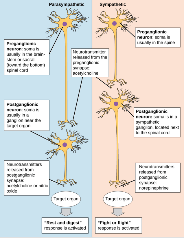
Explanation: The sympathetic chain, also known as the sympathetic trunk, is an important part of the autonomic nervous system. It plays a crucial role in the sympathetic response, which is responsible for the “fight or flight” reaction. The sympathetic chain consists of interconnected ganglia and nerve fibres.
Grey rami communicantes are responsible for carrying postganglionic sympathetic fibres from the sympathetic chain to spinal nerves. These fibres innervate structures in the body, including blood vessels and sweat glands.
White rami communicantes are responsible for carrying preganglionic sympathetic fibres from the spinal cord to the sympathetic ganglia in the chain. These preganglionic fibres originate in the thoracolumbar region of the spinal cord.
Option D is the correct answer because it accurately describes the role of grey and white rami communicantes in the sympathetic chain, which is an essential component of the sympathetic division of the autonomic nervous system.
Option A is incorrect since voluntary muscle movement is controlled by the somatic nervous system, not the sympathetic nervous system which is a branch of the autonomic nervous system.
Option B is incorrect since this is the definition of sensory afferent neurons of the peripheral nervous system.
Option C is incorrect because this is the function of the parasympathetic nervous system. The sympathetic nervous system is responsible for “fight or flight” response.
Option E is incorrect because the sympathetic nervous system carries information from the central nervous system (specifically from the thoracic and lumbar regions of the spinal cord) to various organs and tissues (e.g. muscles & eyes) to prepare it for “fight or flight” responses.
Question 2:
Which neurotransmitter plays a critical role in regulating mood and is often implicated in depression and anxiety disorders?
A) Dopamine
B) Serotonin
C) GABA
D) Glutamate
E) Acetylcholine
Answer: B) Serotonin
Explanation: Serotonin is a neurotransmitter involved in regulating mood, among other functions. Imbalances in serotonin levels are associated with mood disorders.
Option A is incorrect because dopamine plays a key role in reward, motivation, pleasure, and motor control. It helps regulate movement, emotional responses, and the ability to experience pleasure and pain.
Option C is incorrect because GABA is an inhibitory neurotransmitter that’s helps to regulate anxiety, promote relaxation, and maintain a balance in brain activity.
Option D is incorrect since glutamate is an excitatory that plays a key role in movement, learning, memory, and overall brain function.
Option E is incorrect since acetylcholine is mainly involved in smooth muscle contraction.
Question 3:
What is the primary function of the cerebellum in the brain?
A) Memory consolidation
B) Motor coordination and balance
C) Amplification of movement
D) Selecting the correct movement
E) Emotion regulation
Answer: B) Motor coordination and balance
Explanation: The cerebellum is primarily responsible for coordinating voluntary muscle movements and maintaining balance and posture.
Option A is incorrect because memory consolidation primarily involves the hippocampus and certain cortical areas, not the cerebellum.
Option C is incorrect since amplification of movement is regulated by the basal ganglia. Damage to the basal ganglia may cause bradykinesia (limited movement) resulting in Parkinson’s Disease or dyskinesia (excess movement) resulting in Huntington’s Disease.
Option D is incorrect since selecting the correct movement is regulated by the secondary motor cortex in the frontal lobe (e.g. supplementary motor cortex & pre-motor cortex). These regions are involved in planning and coordinating movements. Damage to the secondary motor cortex may cause apraxia (difficulties in planning and executing movements).
Option E is incorrect since emotional regulation involves several areas of the brain, including the limbic system (amygdala and hippocampus) and prefrontal cortex, not the cerebellum.
Question 4:
In which neurological disorder do individuals experience sudden, recurrent, and unprovoked seizures?
A) Huntington’s disease
B) Parkinson’s disease
C) Alzheimer’s disease
D) Epilepsy
E) Multiple sclerosis
Answer: D) Epilepsy
Explanation: Epilepsy is a neurological disorder characterised by recurrent and unprovoked seizures, which result from abnormal electrical activity in the brain.
Option A is incorrect since Huntington’s disease is a neurodegenerative disorder characterised mainly by involuntary movements It does not typically involve sudden, recurrent, and unprovoked seizures.
Option B is incorrect because Parkinson’s disease is a movement disorder characterised by tremors, rigidity, bradykinesia (slowness of movement). It does not involve seizures.
Option C is incorrect because Alzheimer’s disease is a progressive neurodegenerative disorder characterized by memory loss and cognitive decline.
Option E is incorrect because Multiple sclerosis is a demyelinating disorder of the central nervous system that can lead to a wide range of neurological symptoms such as muscle weakness, spasticity & balance & coordination problems, but does not involve seizures.
Question 5:
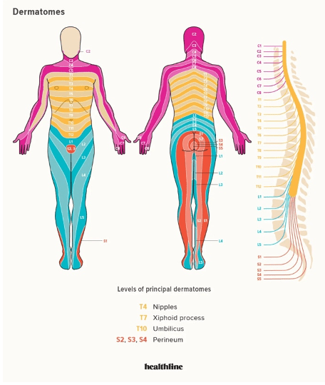
Which of the following accurately describes the function of a dermatome?
A) It is a motor neuron pathway in the limb.
B) It is a region of skin supplied by a single spinal nerve.
C) It is a specialised sensory structure found in the dermis of the skin.
D) It is a specialised sensory receptor.
E) It is a region of skin supplied by multiple spinal nerves.
Answer: B) It is a region of skin supplied by a single spinal nerve.
Explanation: A dermatome is an area of skin supplied by sensory fibers from a single spinal nerve. They are important for assessing sensory function in specific regions of the body.
Option A is incorrect because a dermatome is not a motor neuron pathway but rather a region of skin.
Option C is incorrect because a dermatome is not a specialised sensory structure, but rather a specific region of skin that is innervated by sensory fibres from a single spinal nerve e.g. T10 dermatome supplies the umbilical region.
Option D is incorrect because a dermatome is not a specialised sensory receptor but a region of sensory innervation.
Option E is incorrect because a dermatome is defined by the area of skin supplied by a single spinal nerve, not multiple spinal nerves.
Question 6:
Which neurotransmitter is primarily associated with the reward pathway and is often implicated in addiction?
A) Serotonin
B) GABA
C) Glutamate
D) Dopamine
E) Acetylcholine
Answer: D) Dopamine
Explanation: Dopamine is a neurotransmitter associated with the brain’s reward system and plays a central role in reinforcement and motivation, making it relevant to addiction-related behaviours.
Option A is incorrect because serotonin is primarily involved in mood regulation, REM sleep, and appetite, rather than the reward pathway.
Option B is incorrect because GABA is the main inhibitory neurotransmitter in the brain, involved in reducing neuronal excitation, and not directly associated with the reward pathway or addiction.
Option C is incorrect because glutamate is the main excitatory neurotransmitter in the brain, involved in synaptic plasticity and learning, but it is not primarily associated with the reward pathway or addiction.
Option E is incorrect because acetylcholine plays a role in neuromuscular junctions, autonomic nervous system function, and cognitive processes, but it is not primarily associated with the reward pathway or addiction.
Question 7:
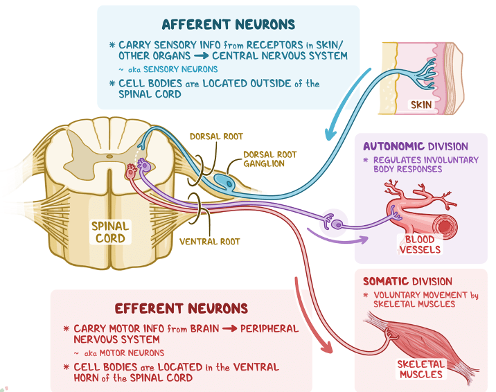
Which type of nerve is responsible for transmitting information from the viscera to the brain?
A) Sensory nerve
B) Motor nerve
C) Afferent nerve
D) Somatic nerve
E) Efferent nerve
Answer: C) Afferent nerve
Explanation: Afferent nerves (also known as sensory nerves) are responsible for transmitting sensory information from the viscera (internal organs) to the brain. These nerves carry signals related to sensations such as pain, pressure, and organ function back to the central nervous system for processing and response.
Option A is less correct than option C because “sensory nerves” generally refer to nerves that carry sensory information from all parts of the body to the CNS, including from the skin and muscles, whereas “afferent nerves” specifically emphasise the function of transmitting sensory signals from internal organs to the CNS.
Option B is incorrect because motor nerves transmit signals from the brain or spinal cord to muscles and glands, controlling movement and glandular secretions, not from the viscera to the brain.
Option D is incorrect because somatic nerves primarily innervate the skin, skeletal muscles, and joints, responsible for conscious sensory perception and voluntary movement, but not specifically transmitting information from the viscera to the brain.
Option E is incorrect because efferent nerves carry signals away from the brain or spinal cord back to muscles, glands or viscera, controlling motor functions, but not specifically involved in transmitting sensory information from the viscera to the brain.
Question 8:
What is the primary function of the neuromuscular junction?
A) Transmit sensory information to the spinal cord.
B) Relay motor commands from the brain to the muscles.
C) Coordinate muscle contractions.
D) Connect pre & postsynaptic neuron.
E) Transmit signals from motor neurons to muscle fibres.
Answer: E) Transmit signals from motor neurons to muscle fibres.
Explanation: The neuromuscular junction is responsible for transmitting signals from motor neurons to muscle fibres, leading to muscle contraction. This process begins with the release of acetylcholine from the motor neuron, which binds to nicotinic receptors on the muscle fibre membrane. This binding event leads to depolarisation of the muscle membrane and an influx of calcium ions into the muscle cell. The increase in intracellular calcium concentration triggers the interaction between actin and myosin filaments, known as the power stroke, which generates muscle movement.
Option A is incorrect because the primary function of the neuromuscular junction is not to transmit sensory information to the spinal cord, but rather to transmit signals from motor neurons to muscle fibres, initiating muscle contraction.
Option B is incorrect because while neuromuscular junctions do relay signals from the nervous system to muscles, they specifically transmit signals from motor neurons in the brain to muscle fibres to elicit muscle contraction, rather than relaying motor commands from the brain to muscles. Option E is a better & more specific definition of a neuromuscular junction.
Option C is incorrect because although neuromuscular junctions contribute to muscle contractions, their primary function is to transmit signals from motor neurons to muscle fibres, initiating muscle contraction, rather than coordinating muscle contractions.
Option D is incorrect because the neuromuscular junction does not connect pre and post-synaptic neuron. Instead, it connects a motor neuron (presynaptic neuron) to a muscle fibre, not a postsynaptic neuron, facilitating the transmission of signals from the neuron to the muscle.

Question 9:
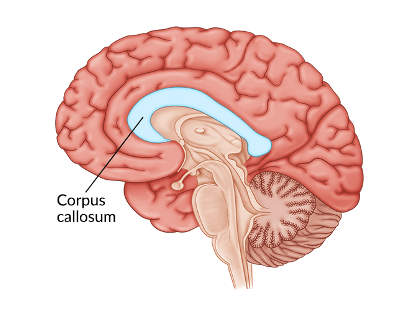
Which structure connects the two hemispheres in the central nervous system?
a) Arbor vitae
b) Thalamus
c) Pons
d) Septum pellucidum
e) Corpus callosum
Answer: e) Corpus callosum
Explanation: The corpus callosum is a large bundle of nerve fibres (called commissural fibres) that connect the left and right cerebral hemispheres. It allows for communication and coordination between the two hemispheres, facilitating integrated brain function.
Option A is incorrect because arbor vitae refers to the tree-like appearance of white matter in the cerebellum, not a structure connecting the hemispheres of the brain.
Option B is incorrect because the thalamus is a central relay station for sensory information to the cerebral cortex and does not connect the two hemispheres of the brain.
Option C is incorrect because the pons is a structure in the brainstem involved in respiration, and relaying information between the cerebrum and the cerebellum, but it does not connect the two hemispheres.
Option D is incorrect because the septum pellucidum is a thin membrane that separates the lateral ventricles of the brain (that form the majority of CSF).
Question 10:
In the context of the autonomic nervous system, what is the primary function of the grey rami communicantes?
A) Transmit sensory information to the spinal cord.
B) Carry postganglionic sympathetic fibres from the sympathetic chain to spinal nerves.
C) Relay motor commands from the brain to the muscles.
D) Connect sensory neurons to the spinal cord.
E) Transmit signals from the brain to the spinal cord.
Answer: B) Carry postganglionic sympathetic fibres from the sympathetic chain to spinal nerves.
Explanation: Grey rami communicantes carry postganglionic sympathetic fibres from the sympathetic chain to spinal nerves, facilitating sympathetic innervation of various body structures, playing a role in regulating involuntary bodily functions like heart rate and digestion.
Option A is incorrect because transmitting sensory information to the spinal cord is typically associated with white communicantes rather than the grey rami communicantes.
Option C is incorrect because relaying motor commands from the brain to the muscles is a function primarily associated with motor neurons in the somatic nervous system, not the autonomic nervous system which is where the grey rami communicantes are found.
Option D is incorrect because connecting sensory neurons to the spinal cord is not a function of the grey rami communicantes. Sensory neurons enter the spinal cord through dorsal root ganglia.
Option E is incorrect because transmitting signals from the brain to the spinal cord is more related to descending pathways involving motor commands rather than the grey rami communicantes in the autonomic nervous system.
Question 11:
Which part of the brainstem is responsible for regulating essential functions such as heart rate, respiration, and blood pressure?
A) Medulla oblongata
B) Midbrain
C) Pons
D) Cerebellum
E) Thalamus
Answer: A) Medulla oblongata
Explanation: The medulla oblongata, located in the brainstem, is responsible for regulating vital autonomic functions, including heart rate, respiration, and blood pressure. It contains the dorsal respiratory group (DRG) and ventral respiratory group (VRG), which are the respiratory centres. Additionally, it has receptors that can detect changes in the pH of the cerebrospinal fluid (CSF) to modulate heart rate and blood pressure accordingly.
Option B is incorrect because the midbrain primarily processes sensory information, coordinates motor responses, and regulates arousal and alertness, but it is not directly responsible for regulating essential functions such as heart rate, respiration, and blood pressure.
Option C is incorrect because while the pons serves as a bridge between different parts of the brain and plays a role in regulating sleep, posture, and some aspects of breathing, it is not primarily responsible for controlling heart rate, respiration, and blood pressure.
Option D is incorrect because the primary function of the thalamus is to coordinate voluntary movements, balance, and motor learning, rather than regulating essential autonomic functions like heart rate, respiration, and blood pressure.
Option E is incorrect because the thalamus acts as a relay station for sensory information to the cerebral cortex and is involved in sensory interpretation.
Question 12:
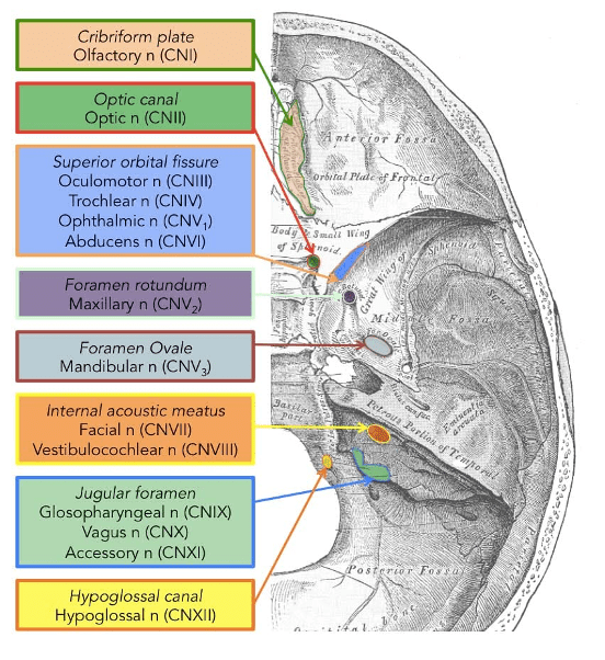
Which cranial nerve is responsible for controlling tongue movement?
A) Cranial nerve X (Vagus nerve)
B) Cranial nerve V (Trigeminal nerve)
C) Cranial nerve VII (Facial nerve)
D) Cranial nerve IX (Glossopharyngeal nerve)
E) Cranial nerve XII (Hypoglossal nerve)
Answer: E) Cranial nerve XII (Hypoglossal nerve)
Explanation: The hypoglossal nerve exits the skull through the hypoglossal canal (foramen) and controls tongue movement.
Option A is incorrect because the vagus nerve (Cranial nerve X) primarily innervates organs in the thorax and abdomen such as the heart & stomach, playing roles in autonomic functions like heart rate, digestion, and respiratory rate regulation. It does not directly control tongue movement.
Option B is incorrect because the trigeminal nerve (Cranial nerve V) is primarily responsible for sensory innervation of the face and motor functions such as chewing. It does not control tongue movement.
Option C is incorrect because the facial nerve (Cranial nerve VII) innervates muscles of facial expression, taste from the anterior two-thirds of the tongue, and secretion of tears and saliva.
Option D is incorrect because the glossopharyngeal nerve (Cranial nerve IX) provides sensory innervation to the oropharynx and taste sensation from the posterior one-third of the tongue, as well as motor control over some muscles in the throat.
Question 13:
What is the primary function of the arachnoid mater in the meninges?
A) To provide cushioning and protection
B) To produce cerebrospinal fluid (CSF)
C) To anchor the brain to the skull
D) To regulate blood flow to the brain
E) To serve as a barrier to infection
Answer: A) To provide cushioning and protection
Explanation: The arachnoid mater, located between the dura mater and pia mater, provides cushioning and protection for the brain and spinal cord.
Option B is incorrect because the production of cerebrospinal fluid (CSF) primarily occurs in the choroid plexus within the ventricles of the brain, not in the arachnoid mater.
Option C is incorrect because the structure responsible for anchoring the brain to the skull is the dura mater, not the arachnoid mater.
Option D is incorrect because the regulation of blood flow to the brain involves mechanisms such as autoregulation by blood vessels and is not the arachnoid mater.
Option E is incorrect because the barrier function against infection is primarily attributed to the blood-brain barrier, rather than the arachnoid mater.

Question 14:
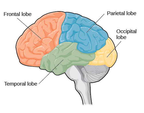
Which major functional area of the cerebral hemispheres is responsible for processing sensory information such as touch and proprioception?
A) Frontal lobe
B) Occipital lobe
C) Parietal lobe
D) Temporal lobe
E) Limbic lobe
Answer: C) Parietal lobe
Explanation: The parietal lobe is primarily responsible for processing sensory information related to touch, temperature, and proprioception.
Option A is incorrect because the frontal lobe is primarily involved in motor control, planning, and decision-making.
Option B is incorrect because the occipital lobe is primarily responsible for processing visual information.
Option D is incorrect because the temporal lobe is mainly associated with auditory processing, language comprehension, and memory functions.
Option E is incorrect because there is no such thing as limbic lobe. Instead, the limbic system is involved in emotions, behaviour, motivation, and memory formation.
Question 15:
What is the primary function of autonomic ganglia?
A) Relay sensory information to the brain.
B) Coordinate voluntary muscle movements.
C) Transmit motor commands from the spinal cord.
D) Regulate involuntary bodily functions.
E) Transmit signals from the brain to the muscles.
Answer: D) Regulate involuntary bodily functions.
Explanation: Autonomic ganglia play a crucial role in regulating involuntary bodily functions, such as heart rate, digestion, and respiratory rate.
Option A is incorrect because autonomic ganglia relay motor commands to viscera, glands & tissue rather than sensory information to the brain. They are part of the autonomic nervous system, which controls involuntary bodily functions such as heart rate, digestion, and respiratory rate.
Option B is incorrect because autonomic ganglia do not coordinate voluntary muscle movements. Voluntary muscle movements are primarily controlled by the somatic nervous system, which includes motor neurons that innervate skeletal muscles.
Option C is incorrect because autonomic ganglia do not transmit motor commands from the spinal cord. Instead, they receive preganglionic fibres from the spinal cord or brainstem and transmit postganglionic fibres to target organs to regulate autonomic functions.
Option E is incorrect because autonomic ganglia do not transmit signals directly from the brain to muscles. They mediate signals between preganglionic neurons from the central nervous system and postganglionic neurons that innervate smooth muscle, cardiac muscle, and glands.
Question 16:
What is the primary role of neurons whose cell bodies are in the dorsal root ganglion?
A) Transmit motor commands to muscles.
B) Relay sensory information from central nervous system to muscles.
C) Relay sensory information to the central nervous system.
D) Regulate autonomic functions.
E) Relay motor impulses from the central nervous system to muscles.
Answer: C) Relay sensory information to the central nervous system.
Explanation: The dorsal root ganglion (DRG) contains the cell bodies of sensory neurons that receive information from sensory receptors in the periphery, such as skin, muscles, and joints. These sensory neurons detect touch, temperature, pain, and proprioception and transmit this information to the central nervous system for processing.
Option A is incorrect because neurons in the DRG primarily transmit sensory information to the CNS rather than motor commands to muscles.
Option B is incorrect because neurons in the DRG relay sensory information from the periphery (like skin, muscles, and joints) to the central nervous system (spinal cord and brain
Option D is incorrect because neurons in the dorsal root ganglion do not regulate autonomic functions. Autonomic functions, such as heart rate, digestion, and respiration, are primarily regulated by neurons in autonomic ganglia and other parts of the autonomic nervous system.
Option E is incorrect because neurons in the dorsal root ganglion do not relay motor impulses from the central nervous system to muscles. Motor impulses are transmitted by motor neurons located in the spinal cord and brainstem, which project their axons directly to muscles to control voluntary movements.
Question 17:
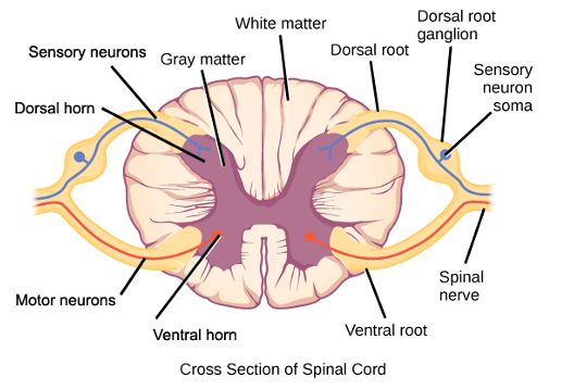
In the peripheral nervous system, where are the cell bodies of motor neurons typically located?
A) In the ventral horn of the spinal cord.
B) In the dorsal root ganglion.
C) In the dorsal horn of the spinal cord
D) In the brainstem.
E) In the lateral horn of the spinal cord
Answer: A) In the ventral horn of the spinal cord.
Explanation: The cell bodies of motor neurons in the peripheral nervous system are typically found in the ventral horn of the spinal cord. These motor neurons control voluntary muscle movements.
Option B is incorrect because the dorsal root ganglion (DRG) in the peripheral nervous system contains the cell bodies of sensory neurons, not motor neurons.
Option C is incorrect because the dorsal horn of the spinal cord primarily contains axons of sensory neurons involved in transmitting sensory information to the brain.
Option D is incorrect because the brainstem contains nuclei involved in controlling vital functions such as respiration, heart rate, and digestion, but not cell bodies of motor neurons.
Option E is incorrect because the lateral horn of the spinal cord contains cell bodies of autonomic motor neurons involved in regulating involuntary functions of organs, glands, and smooth muscle, not skeletal muscles.
Question 18:
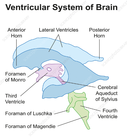
Which structure connects the third and fourth ventricles in the brain, allowing cerebrospinal fluid (CSF) to flow between them?
A) Cerebral aqueduct (Sylvian aqueduct)
B) Interventricular foramen (Foramen of Monro)
C) Lateral aperture (Foramen of Luschka)
D) Median aperture (Foramen of Magendie)
E) Septum pellucidum
Answer: A) Cerebral aqueduct (Sylvian aqueduct)
Explanation: The cerebral aqueduct connects the third and fourth ventricles, facilitating the flow of cerebrospinal fluid (CSF).
Option B is incorrect because the interventricular foramen (Foramen of Monro) connects the lateral ventricle to the third ventricle.
Option C is incorrect because the lateral aperture (Foramen of Luschka) is located in the lateral recesses of the fourth ventricle and allows CSF to exit the ventricular system into the subarachnoid space surrounding the brain.
Option D is incorrect because the median aperture (Foramen of Magendie) is a midline opening in the fourth ventricle which allows CSF to exit the ventricular system into the subarachnoid space.
Option E is incorrect because the septum pellucidum is a thin membrane located between the lateral ventricles of the brain. It does not connect the third and fourth ventricles or play a role in CSF circulation between these structures.
Question 19:
From which part of the brainstem do cranial nerves 5 through 8 (CN V-VIII) arise?
A) Medulla oblongata
B) Midbrain
C) Pons
D) Cerebellum
E) Cerebrum
Answer: C) Pons
Explanation: Cranial nerves 5 through 8, which include the trigeminal (CN V), abducens (CN VI), facial (CN VII), and vestibulocochlear (CN VIII) nerves, arise from the pons, which is one of the three main divisions of the brainstem.
Option A is incorrect because the medulla oblongata primarily gives rise to cranial nerves 9 through 12 (CN IX-XII), namely the glossopharyngeal nerve (IX), vagus nerve (X), accessory nerve (XI), and hypoglossal nerve (XII).
Option B is incorrect because the midbrain, also known as the mesencephalon, gives rise to cranial nerves 3 and 4 (CN III and IV). Cranial nerve III (oculomotor nerve) & cranial nerve IV (trochlear nerve).
Option D is incorrect because the cerebellum is not a part of the brainstem & does not give rise to any cranial nerves.
Option E is incorrect because the cerebrum gives rise to cranial nerves I & II; the olfactory (CNI) & optic nerve (CNII).
Question 20:
Which part of the brain is responsible for processing visual information and is located in the posterior part of the cerebral hemispheres?
A) Frontal lobe
B) Occipital lobe
C) Parietal lobe
D) Temporal lobe
E) Insular cortex
Answer: B) Occipital lobe
Explanation: The occipital lobe is primarily responsible for processing visual information and is located in the posterior part of the cerebral hemispheres.
Option A is incorrect because the frontal lobe of the brain is primarily responsible for motor control, decision making, problem-solving, and speech production.
Option C is incorrect because the parietal lobe is involved in sensory processing, spatial awareness, and attention. It integrates sensory information from various parts of the body and helps in spatial orientation.
Option D is incorrect because the temporal lobe is involved in auditory processing, memory, and language comprehension. It is located on the sides of the brain and plays a key role in auditory perception and speech.
Option E is incorrect because the insular cortex is a small region located deep within the lateral sulcus of the brain. It is involved in functions related to emotions, empathy, and autonomic control.
Question 21:
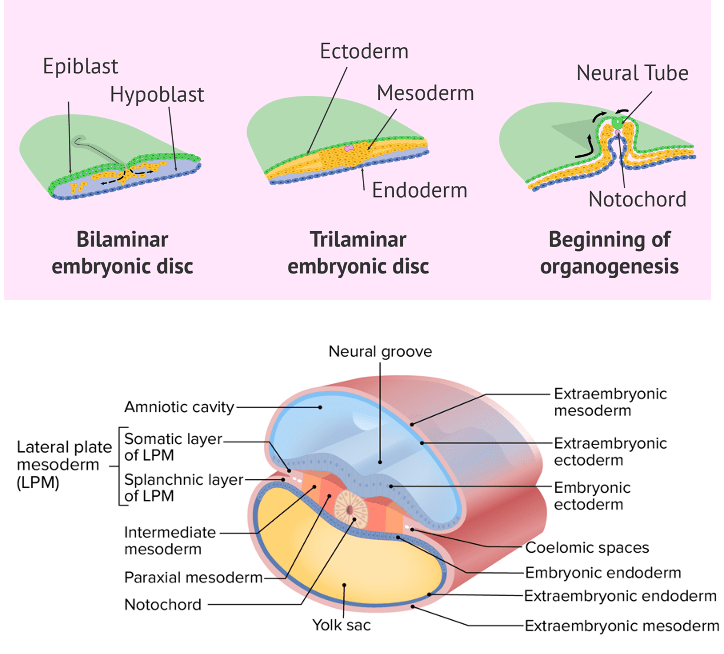
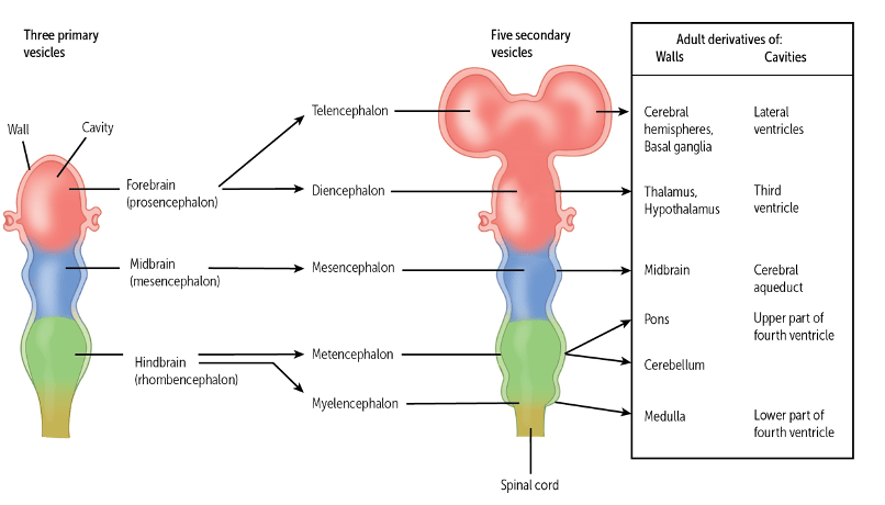
During embryonic development, the neural tube forms from which germ layer?
A) Mesoderm
B) Endoderm
C) Ectoderm
D) Somites
E) Lateral plate mesoderm
Answer: C) Ectoderm
Explanation: The neural tube forms from the ectodermal layer during the early stages of embryonic development. The ectoderm is the outermost germ layer during embryonic development and gives rise to structures such as the nervous system (including the neural tube), skin, and hair.
Option A is incorrect because the mesoderm primarily develops into structures such as muscles, bones, and the circulatory system.
Option B is incorrect because the endoderm develops into the lining of organs such as the gastrointestinal tract and respiratory system.
Option D is incorrect because somites are derived from paraxial mesoderm and gives rise to structures such as skeletal muscle, bones, and dermis.
Option E is incorrect because the lateral plate mesoderm develops into the circulatory system, connective tissues, and certain organs such as the spleen and kidneys.
Question 22:
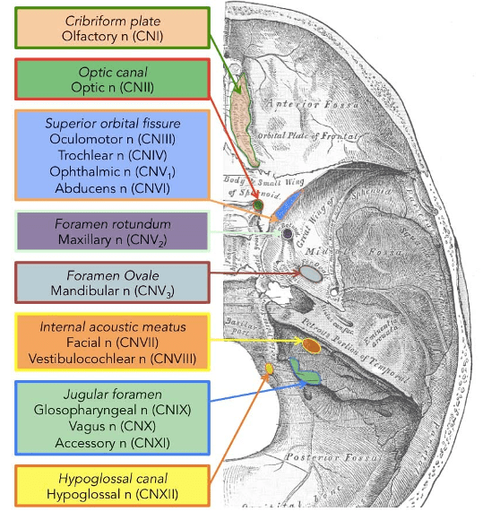
Which cranial nerve exits the skull through the Jugular foramen and is responsible for controlling vital functions such as respiration and heart rate?
A) Cranial nerve IX (Glossopharyngeal nerve)
B) Cranial nerve X (Vagus nerve)
C) Cranial nerve XI (Accessory nerve)
D) Cranial nerve XII (Hypoglossal nerve)
E) Cranial nerve VIII (Vestibulocochlear nerve)
Answer: B) Cranial nerve X (Vagus nerve)
Explanation: The vagus nerve exits the skull through the foramen magnum and is responsible for controlling vital functions such as respiration and heart rate.
Cranial nerves IX, X & XI all exit the skull via the jugular foramen however options A & C are incorrect because CN IX (Glossopharyngeal nerve) is responsible for taste sensation in the posterior one-third of the tongue, swallowing, and secretion of saliva. CN XI (Accessory nerve) is responsible for innervating trapezius & sternocleidomastoid muscles of the neck & back to initiate neck movement.
Option D is incorrect because cranial nerve XII, the Hypoglossal nerve, exits through the Hypoglossal canal and is responsible for motor control of the muscles of the tongue.
Option E is incorrect because cranial nerve VIII, the Vestibulocochlear nerve, exits through the internal acoustic meatus and is responsible for transmitting sensory information related to hearing and balance.
Question 23:
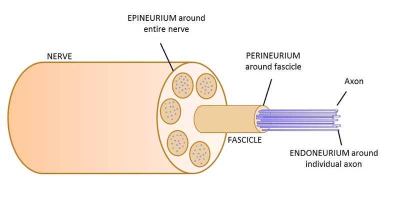
What is the connective tissue component that surrounds individual axons within a nerve bundle?
A) Epineurium
B) Perineurium
C) Endoneurium
D) Myelin sheath
E) Fascicle
Answer: C) Endoneurium
Explanation: The endoneurium is the connective tissue component that surrounds individual axons within a nerve bundle. It provides support and protection to the axons as they travel within a nerve.
Option A is incorrect because the epineurium surrounds the entire nerve bundle, providing protection and support to the entire nerve.
Option B is incorrect because the perineurium surrounds bundles of axons, known as fascicles, within a nerve.
Option D is incorrect because the myelin sheath surrounds some axons, not all such as the axons of sensory & motor neurons, but not relay neurones to facilitate faster nerve conduction.
Option E is incorrect because a fascicle refers to a bundle of axons surrounded by the perineurium within a nerve.
Question 24:
Which structure in the skull allows the passage of cranial nerves III, IV, V1, and VI?
A) Foramen ovale
B) Foramen spinosum
C) Superior orbital fissure
D) Foramen lacerum
E) Foramen rotundum
Answer: E) Superior orbital fissure
Explanation: The superior orbital fissure allows cranial nerves III (oculomotor), IV (trochlear), and VI (abducens) to exit the skull and supply the muscles of the eye.
Option A is incorrect because the foramen ovale allows the passage of cranial nerve V3 (mandibular nerve).
Option B is incorrect because the foramen spinosum permits the passage of the middle meningeal artery, middle meningeal vein, and the meningeal branch of the mandibular nerve.
Option D is incorrect because foramen lacerum transmits small arteries and veins, not cranial nerves.
Option E is incorrect because foramen rotundum transmits the maxillary nerve (cranial nerve V2).
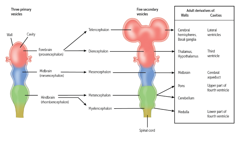
Question 25:
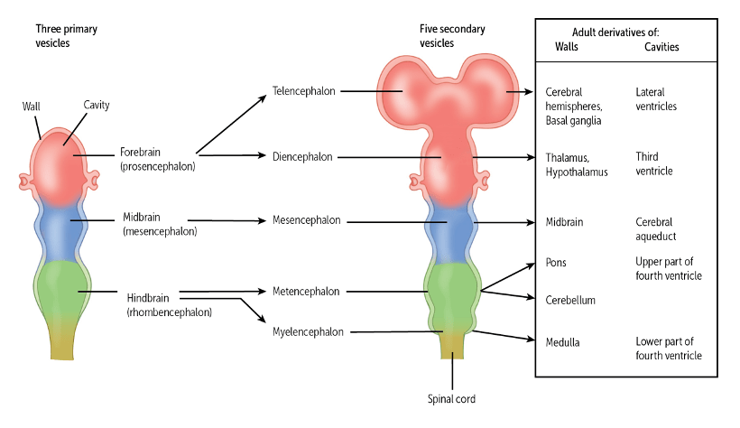
Which brain vesicle primarily gives rise to the adult cerebrum?
A) Prosencephalon (forebrain)
B) Mesencephalon (midbrain)
C) Rhombencephalon (hindbrain)
D) Diencephalon
E) Telencephalon
Answer: E) Telencephalon
Explanation: The telencephalon, a subdivision of the prosencephalon (forebrain), gives rise to the adult cerebrum.
Although option A is correct, the prosencephalon gives rise to the diencephalon & telencephalon. The telencephalon ultimately becomes the cerebrum which is the more correct answer.
Option B is incorrect because the mesencephalon develops into the midbrain.
Option C is incorrect because the rhombencephalon (hindbrain), gives rise to the metencephalon (which gives rise to pons & cerebellum) & myelencephalon (which gives rise to the medulla oblongata).
Option D is incorrect because the diencephalon forms structures like the thalamus and hypothalamus.

Question 26:
What is the embryonic precursor of the medulla oblongata in the adult brain?
A) Metencephalon
B) Myelencephalon
C) Diencephalon
D) Telencephalon
E) Mesencephalon
Answer: B) Myelencephalon
Explanation: The myelencephalon is the embryonic precursor of the medulla oblongata in the adult brain.
Option A is incorrect because the metencephalon gives rise to the pons and cerebellum.
Option C is incorrect because the diencephalon forms structures like the thalamus and hypothalamus.
Option D is incorrect because the telencephalon develops into the cerebrum, not the medulla oblongata.
Option E is incorrect because the mesencephalon gives rise to the midbrain.
Question 27:
Which of the following does NOT originate from the ectoderm during embryonic development?
A) Skin
B) Nervous system
C) Muscles
E) Hair
E) Nails
Answer: C) Muscles
Explanation: Muscles primarily originate from the mesoderm during embryonic development, not the ectoderm.
Options A, D & E are all derived from surface ectoderm whereas the option B, the nervous system is derived from neuroectoderm (specifically the neural tube).
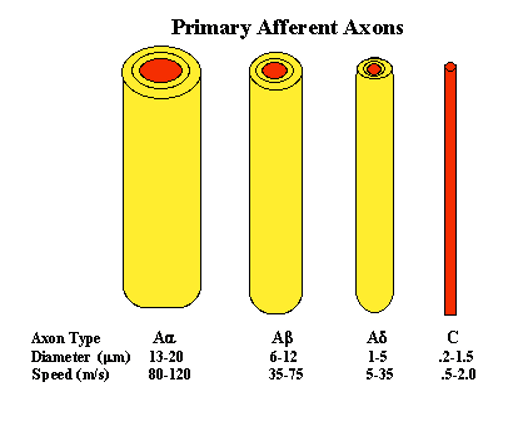
Question 28:
C fibres are primarily responsible for transmitting which type of sensory information?
A) Sharp, localised pain
B) Warm temperature and slow pain
C) Muscle proprioception
D) Fast, voluntary muscle contractions
E) Fine touch and vibration
Answer: B) Warm temperature and slow pain
Explanation: C fibres are unmyelinated and slow-conducting fibres that transmit sensory information related to warm temperature and pain.
Option A is incorrect because sharp & localised pain is transmitted via Aδ fibres (delta).
Option C is incorrect because muscle proprioception is detected by type Ia fibres.
Option D is incorrect because fast, voluntary muscle contractions are transmitted by alpha motor fibres.
Option E is incorrect fine touch & vibration is transmitted by Aβ fibres.
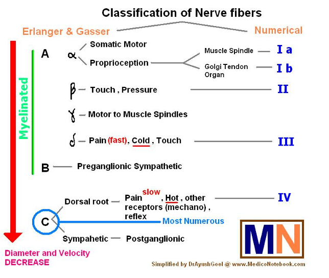
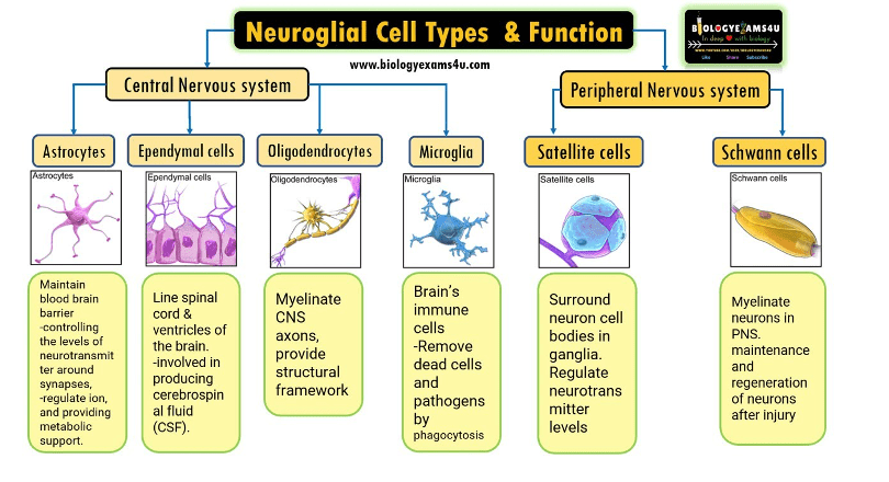
Question 29:
What is the primary function of Schwann cells in the nervous system?
A) Formation of myelin sheaths in the central nervous system
B) Formation of myelin sheaths in the peripheral nervous system
C) Synthesising cerebrospinal fluid
D) Maintaining the blood-brain barrier
E) Regulating immunity in the nervous system
Answer: B) Formation of myelin sheaths in the peripheral nervous system
Explanation: Schwann cells are responsible for forming myelin sheaths around axons in the peripheral nervous system, facilitating faster signal transmission whilst oligodendrocytes are responsible for myelin sheath in CNS.
Option A is incorrect because oligodendrocytes form myelin sheaths in the central nervous system.
Option C is incorrect because ependymal cells synthesise cerebrospinal fluid.
Option D is incorrect because astrocytes are primarily involved in maintain the blood brain barrier.
Option E is incorrect because microglial cells are involved in regulating immunity in the nervous system by phagocytosing pathogens.

Question 30:
A-alpha fibres are primarily associated with which physiological function?
A) Proprioception
B) Carrying motor signals to skeletal muscles to initiate their contraction
C) Sensing changes in temperature
D) Transmitting touch sensations
E) Cause contraction of muscle spindles
Answer: B) Carrying motor signals to skeletal muscles
Explanation: A-alpha fibres are large, myelinated fibres that primarily carry motor signals to skeletal muscles, allowing for fast and efficient voluntary muscle contractions.
Option A is incorrect because proprioception is primarily associated with Ia fibres, which transmit sensory information from muscle providing feedback about muscle length and tension.
Option C is incorrect because temperature sensation is primarily mediated by A-delta and C fibres.
Option D is incorrect because A-alpha fibres transmit motor impulses to skeletal muscle not sensory information such as touch sensations.
Option E is incorrect because contraction of muscle spindles is transmitted by gamma motor neurons which is more specific than alpha motor neuron.
Question 31:
What is the primary function of astrocytes in the nervous system?
A) Phagocytosis of pathogens and debris
B) Formation of myelin sheaths
C) Providing structural support and maintaining the blood-brain barrier
D) Regulation of neurotransmitter release
E) Initiating immune responses
Answer: C) Providing structural support and maintaining the blood-brain barrier
Explanation: Astrocytes provide structural support to neurons and help maintain the integrity of the blood-brain barrier in the central nervous system (CNS).
Option A is incorrect because microglial cells are primarily responsible for phagocytosis of pathogens and debris in the nervous system.
Option B is incorrect because oligodendrocytes (in the central nervous system) and Schwann cells (in the peripheral nervous system) are responsible for the formation of myelin sheaths around axons.
Option D is correct. Yes astrocytes do regulate neurotransmitter release, however their main primary function is maintaining the blood-brain barrier.
Option E is incorrect because microglial cells are the resident immune cells of the central nervous system and are primarily involved in initiating immune responses in the nervous system.
Question 32:
During brain development, which structure differentiates into the thalamus, hypothalamus, and epithalamus?
A) Telencephalon
B) Diencephalon
C) Mesencephalon
D) Metencephalon
E) Myelencephalon
Answer: B) Diencephalon
Explanation: The diencephalon differentiates into structures such as the thalamus, hypothalamus, and epithalamus in the adult brain.
Option A is incorrect because the telencephalon differentiates into the cerebral cortex, basal ganglia, and olfactory bulb.
Option C is incorrect because the mesencephalon develops into the midbrain.
Option D is incorrect because the metencephalon develops into the pons and the cerebellum.
Option E is incorrect because the myelencephalon differentiates into the medulla oblongata.
Question 33:
What is the primary function of the blood-brain barrier in the nervous system?
A) Promoting the entry of immune cells into the brain
B) Preventing the passage of nutrients into the brain
C) Protecting the brain from harmful substances and pathogens in the blood
D) Facilitating the exchange of neurotransmitters
E) Regulating blood pressure in the brain
Answer: C) Protecting the brain from harmful substances and pathogens in the blood.
Explanation: The blood-brain barrier serves to protect the brain by limiting the entry of potentially harmful substances and pathogens from the bloodstream.
Option A is incorrect because the blood-brain barrier (BBB) does not promote the entry of immune cells into the brain. In fact, it restricts the entry of immune cells to protect the brain from potential inflammation and damage.
Option B is incorrect because the BBB does not prevent the passage of nutrients into the brain. It selectively allows essential nutrients to pass through while blocking harmful substances.
Option D is incorrect because the BBB does not facilitate the exchange of neurotransmitters. Neurotransmitter exchange occurs at synapses, not through the BBB.
Option E is incorrect because the primary function of the BBB is not to regulate blood pressure in the brain. Blood pressure regulation is managed by other mechanisms within the cardiovascular system.
Question 34:
A-gamma fibers are primarily associated with which physiological function?
A) Transmitting pain signals
B) Carrying motor signals to skeletal muscles
C) Sensory control of muscle spindles
D) Transmitting touch sensations
E) Motor control of muscle spindle
Answer: E) Motor control of muscle spindle
Explanation: A-gamma fibres are primarily associated with motor control of muscle spindles, adjusting the sensitivity of muscle spindles to stretch and ensuring proper muscle function and tone.
Option A is incorrect because transmitting pain signals is primarily the function of C fibres (which transmit chronic pain) and A-delta fibres (which transmit sharp/quick pain).
Option B is incorrect because carrying motor signals to skeletal muscles is primarily the function of A-alpha fibres. Although gamma fibres are a branch of A-alpha fibres meaning that they do carry motor signals to skeletal muscle, their specific function is to carry motor impulses to the spindles of the muscle which is a more correct answer.
Option C is incorrect because the sensory control of muscle spindles is primarily the function of A-beta fibres.
Option D is incorrect because transmitting touch sensations is primarily the function of A-beta fibres.
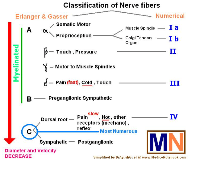
Question 35:
Which type of nerve fibres are primarily associated with fast signal transduction in the nervous system?
A) Myelinated fibres
B) Unmyelinated fibres
C) C fibres
D) A-delta fibres
E) A-alpha fibres
Answer: E) A-alpha fibres
Explanation: A-alpha fibres are myelinated and are primarily associated with the fastest signal transduction in the nervous system, carrying motor signals to skeletal muscles and sensory information related to proprioception.
Myelinated nerve fibres are known for their ability to conduct signals rapidly due to the presence of the myelin sheath, which insulates and speeds up the transmission of electrical impulses along the nerve axon.
Option A is incorrect because while myelinated fibres do facilitate fast signal transduction, this option is too broad and not specific to the type of fibre primarily associated with the fastest conduction.
Option B is incorrect because unmyelinated fibres conduct signals more slowly compared to myelinated fibres. This is due to a lack of saltatory conduction.
Option C is incorrect because C fibres are unmyelinated and are associated with slower signal transmission, involved in transmitting slow pain and temperature signals.
Option D is incorrect because A-delta fibres, while myelinated and faster than C fibres, are not as fast as A-alpha fibres and are involved in transmitting fast pain and temperature signals.
Question 36:
Which supporting cells in the central nervous system (CNS) are responsible for forming myelin sheaths around axons?
A) Oligodendrocytes
B) Astrocytes
C) Microglia
D) Ependymal cells
E) Schwann cells
Answer: A) Oligodendrocytes
Explanation: Oligodendrocytes are responsible for forming myelin sheaths around axons in the central nervous system (CNS). Schwann cells do the same but for the peripheral nervous system (PNS).
Option B is incorrect because astrocytes provide structural support, maintain the blood-brain barrier, and regulate neurotransmitter levels, but they do not form myelin sheaths around axons.
Option C is incorrect because microglia are involved in immune responses in the CNS by acting as macrophages to phagocytose pathogens and debris.
Option D is incorrect because ependymal cells line the ventricles of the brain and the central canal of the spinal cord, and are involved in producing and circulating cerebrospinal fluid (CSF).
Question 37:
In the cross-sectional organization of the spinal cord, which region contains motor neurons that send signals to skeletal muscles?
A) Dorsal horn
B) Lateral horn
C) Ventral horn
D) Central canal
E) Spinal ganglion
Answer: C) Ventral horn
Explanation: The ventral horn of the spinal cord contains motor neurons that send signals to skeletal muscles.
Option A is incorrect because the dorsal horn primarily contains sensory neurons that receive and process incoming sensory information from the peripheral nerves.
Option B is incorrect because the lateral horn contains neurons involved in autonomic (involuntary) functions, specifically those related to the sympathetic nervous system.
Option D is incorrect because the central canal is a cerebrospinal fluid-filled space that runs longitudinally through the length of the entire spinal cord and does not contain motor neurons.
Option E is incorrect because the spinal ganglion, also known as the dorsal root ganglion, contains the cell bodies of sensory neurons that transmit sensory information to the spinal cord, not motor neurons.
Question 38:
At the neuromuscular junction, which neurotransmitter is responsible for transmitting signals from motor neurons to muscle cells?
A) Gamma-aminobutyric acid (GABA)
B) Glycine
C) Acetylcholine
D) Substance P
E) Glutamate
Answer: C) Acetylcholine
Explanation: Acetylcholine is the neurotransmitter responsible for transmitting signals at the neuromuscular junction between motor neurons and muscle cells.
Option A is incorrect because GABA is an inhibitory neurotransmitter in the central nervous system, involved in reducing neuronal excitability.
Option B is incorrect because glycine is an inhibitory neurotransmitter in the central nervous system, particularly in the spinal cord, brainstem, and retina, where it contributes to the processing of motor and sensory information.
Option D is incorrect because Substance P is a neuropeptide involved in the transmission of pain and other sensory information within the central nervous system and the peripheral nervous system.
Option E is incorrect because glutamate is the primary excitatory neurotransmitter in the central nervous system, involved in cognitive functions such as learning and memory. However, it is not the neurotransmitter used at the neuromuscular junction.
Question 39:
Which of the following is not a secondary brain vesicle?
A) Prosencephalon
B) Myelencephalon
C) Metencephalon
D) Diencephalon
E) Telencephalon
Answer: a) Prosencephalon
Explanation: Prosencephalon is the only primary brain vesicle in this list. The prosencephalon gives rise to the telencephalon & diencephalon (which are secondary brain vesicles). Primary brain vesicles develop in the 4th week of embryonic development whereas secondary brain vesicles develop in the 5th week of embryonic development.
Question 40:
In the sympathetic division of the autonomic nervous system, what distinguishes the length of pre-ganglionic and post-ganglionic neurons?
A) Both pre-ganglionic and post-ganglionic neurons have short axons.
B) Both pre-ganglionic and post-ganglionic neurons have long axons.
C) Pre-ganglionic neurons have long axons, while post-ganglionic neurons have short axons.
D) Pre-ganglionic neurons have short axons, while post-ganglionic neurons have long axons.
E) The lengths of axons in the sympathetic division vary widely and are not consistent.
Answer: D) Pre-ganglionic neurons have short axons, while post-ganglionic neurons have long axons.
Explanation: In the sympathetic division of the autonomic nervous system, pre-ganglionic neurons have short axons, typically extending only from the spinal cord to the nearby sympathetic ganglia, while post-ganglionic neurons have longer axons that reach their target organs and tissues. This distinction allows for a two-stage pathway for sympathetic responses, with the ganglia serving as relay points between the central nervous system and the peripheral target organs.
Option C is the length of neurons in the parasympathetic nervous system.

Question 41:
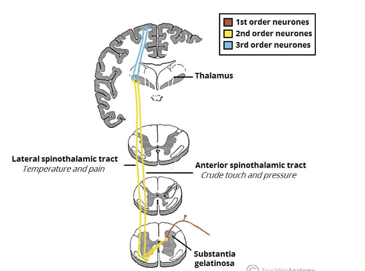
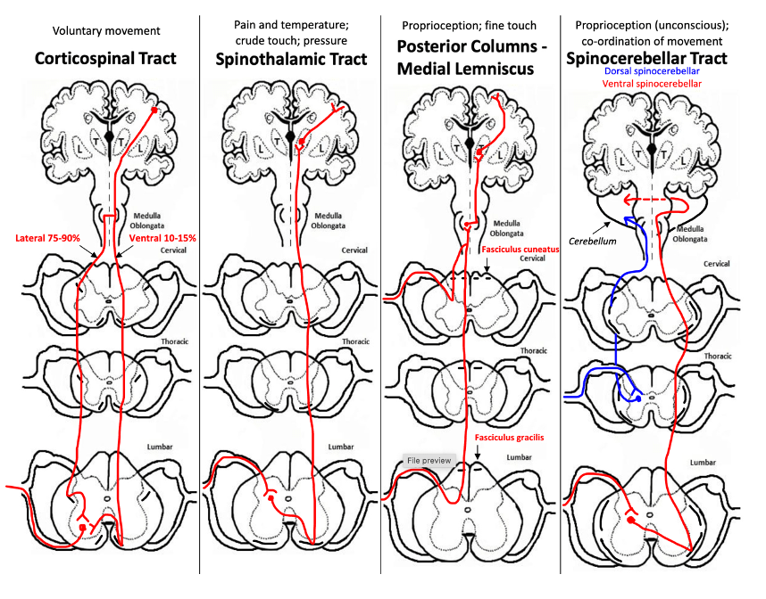
Where does the decussation (crossing over) of fibres of the spinothalamic system occur in the somatosensory pathways?
A) In the dorsal column of the level of entry
B) In the spinal cord at the level of entry
C) In the dorsal root ganglia at the level
D) In the medulla
E) In the thalamus
Answer: B) In the spinal cord at the level of entry
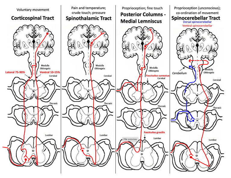
Explanation: The decussation of fibres of the spinothalamic system occurs in the spinal cord, allowing sensory information to cross to the opposite side of the body at the level of entry or 1 or 2 above.
Option A is incorrect because no tract decussates in the dorsal column. 1st order neurons in the dorsal column medial lemniscus tract do indeed ascend in dorsal columns but their 2ndorder neurons decussate in the medulla oblongata.
Option C is incorrect because the dorsal root ganglia do not participate in the decussation of any fibres; they contain the cell bodies of sensory neurons.
Option D is incorrect because while decussation does occur in the medulla for some pathways, such as the dorsal column-medial lemniscus pathway and corticospinal tract, it is not where the spinothalamic tract decussates.
Option E is incorrect because the thalamus is where sensory information is relayed after initial processing and decussation has already occurred.
Question 42:
Which cranial nerve is primarily responsible for controlling the lateral rectus muscle of the eye, which moves the eye laterally?
A) Optic nerve (cranial nerve II)
B) Oculomotor nerve (cranial nerve III)
C) Abducens nerve (cranial nerve VI)
D) Trochlear nerve (cranial nerve IV)
E) Facial nerve (cranial nerve VII)
Answer: C) Abducens nerve (cranial nerve VI)
Explanation: The abducens nerve innervates the lateral rectus muscle, responsible for moving the eye laterally.
Option A is incorrect because the optic nerve is responsible for transmitting visual information from the retina to the brain. It does not control any eye muscles.
Option B is incorrect because while the oculomotor nerve controls most of the muscles that move the eye, it primarily innervates the superior, inferior, and medial rectus muscles, as well as the inferior oblique muscle, not the lateral rectus muscle.
Option D is incorrect because the trochlear nerve innervates the superior oblique muscle, which helps in downward and medial rotation of the eye.
Option E is incorrect because the facial nerve is responsible for controlling the muscles of facial expression, taste sensation from the anterior two-thirds of the tongue, and other functions related to the face, but it does not control any of the extraocular muscles.
Question 43:
What role does the thalamus play in the somatosensory pathway?
A) It serves as the input to the cerebellum.
B) It is responsible for proprioception.
C) It relays sensory information to the cerebral cortex.
D) It controls reflex withdrawal to pain.
E) It houses the sensory homunculus.
Answer: C) It relays sensory information to the cerebral cortex.
Explanation: The thalamus acts as a relay station in the somatosensory pathway, transmitting sensory information to the cerebral cortex for further processing and perception.
Option A is incorrect because the thalamus does not serve as the input to the cerebellum in the somatosensory pathway. Instead, the cerebellum relays impulses to the thalamus, specifically to the ventral lateral nucleus (VL) and the ventral anterior nucleus (VA) of the thalamus.
Option B is incorrect because proprioceptive information is processed in the somatosensory cortex. The thalamus specifically relays sensory information to the cerebral cortex rather than being responsible for proprioception itself.
Option D is incorrect because reflex withdrawal to pain is primarily mediated at the spinal cord level, not in the thalamus.
Option E is incorrect because the sensory homunculus is a representation of the body in the somatosensory cortex, not in the thalamus.
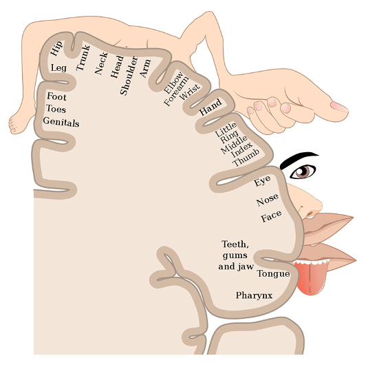
Question 44:
What is the primary role of the sensory homunculus located in the primary somatosensory cortex?
A) Relays sensory information to the somatosensory cortex
B) Relay sensory information to the spinal cord
C) Represent the somatotopic map of the body
D) Regulate autonomic functions
E) Process visual information
Answer: C) Represent the somatotopic map of the body
Explanation: The sensory homunculus is a representation of the somatotopic map of the body in the primary somatosensory cortex, where different areas correspond to sensory input from different parts of the body.
Option A is incorrect because the thalamus relays sensory information to the somatosensory cortex.
Option B is incorrect because the sensory homunculus does not relay sensory information to the spinal cord. Sensory information is first processed in peripheral sensory receptors, transmitted through the spinal cord and brainstem nuclei, and then reaches the somatosensory cortex.
Option D is incorrect because autonomic functions such as heart rate and digestion are primarily controlled by brainstem nuclei and hypothalamic regions, not the somatosensory cortex.
Option E is incorrect because visual information is processed in the visual cortex, located in the occipital lobe of the brain.
Question 45:
Which cranial nerve is responsible for controlling the inferior oblique muscle of the eye?
A) Optic nerve (cranial nerve II)
B) Oculomotor nerve (cranial nerve III)
C) Abducens nerve (cranial nerve VI)
D) Trochlear nerve (cranial nerve IV)
E) Facial nerve (cranial nerve VII)
Answer: B) Oculomotor nerve (cranial nerve III)
Explanation: The oculomotor nerve innervates the inferior oblique muscle of the eye. It also innervates the superior, medial & inferior recti muscles.
Option A is incorrect because the optic nerve (cranial nerve II) is responsible for transmitting visual information from the retina to the brain and does not control any of the extraocular muscles.
Option C is incorrect because the abducens nerve (cranial nerve VI) primarily innervates the lateral rectus muscle, which is responsible for abduction of the eye.
Option D is incorrect because the trochlear nerve (cranial nerve IV) innervates the superior oblique muscle, which is responsible for downward and medial rotation of the eye.
Option E is incorrect because the facial nerve (cranial nerve VII) is responsible for controlling the muscles of facial expression, taste sensation from the anterior two-thirds of the tongue and innervates the salivary glands. It does not control any of the extraocular muscles.
Question 46:
Which neural pathway is primarily responsible for transmitting crude touch sensations to the brain?
A) Dorsal column medial lemniscus pathway
B) Anterior pathway of spinothalamic tract
C) Posterior pathway of spinothalamic tract
D) Lateral corticospinal tract
E) Ventral corticospinal tract
Answer: B) Anterior pathway of spinothalamic tract
Explanation: The anterior pathway of the spinothalamic tract is primarily responsible for transmitting crude touch sensations to the brain. This pathway carries sensory information related to light or crude touch and pressure.
Option A is incorrect because the dorsal column medial lemniscus tract transmits fine touch sensation.
Option C is incorrect because the posterior spinothalamic tract transmits pain and temperature sensations, not crude touch.
Option D is incorrect because the lateral corticospinal tract is involved in motor innervation of distal skeletal muscles (such as fingers) enabling fine movements to be carried out.
Option E is incorrect because the ventral corticospinal tract is also involved in in motor proximal muscles, like those of the trunk.
Question 47:
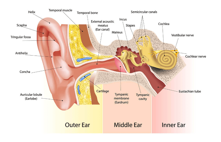
Which part of the ear is responsible for transmitting sound waves from the external ear to the middle ear?
A) Tympanic membrane (eardrum)
B) Pharyngotympanic tube
C) Oval window
D) Auditory ossicles
E) Semicircular canals
Answer: A) Tympanic membrane (eardrum)
Explanation: The tympanic membrane, or eardrum, transmits sound waves/vibrations from the external ear to the middle ear.
Option B is incorrect because the pharyngotympanic tube connects the middle ear to the nasopharynx and helps equalise pressure, but it does not transmit sound waves.
Option C is incorrect because the oval window is part of the middle ear and transmits vibrations from the middle ear to the inner ear, not from the external ear to the middle ear.
Option D is incorrect because the auditory ossicles (malleus, incus, stapes) are responsible for transmitting and amplifying vibrations from the tympanic membrane (eardrum) to the oval window, leading into the inner ear.
Option E is incorrect because the semicircular canals are part of the inner ear and are involved in balance and detecting head movements, not in transmitting sound waves from the external ear to the middle ear.
Question 48:
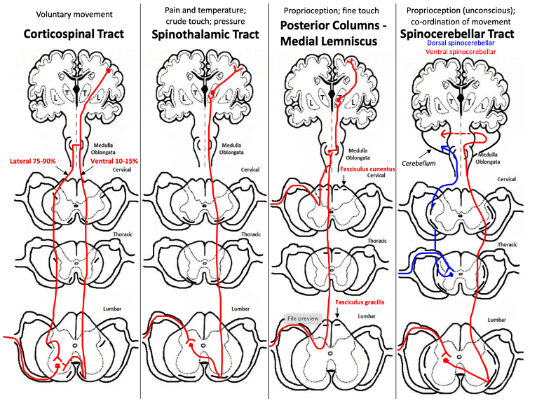
Which of the following sensory modalities is primarily transmitted through the spinocerebellar pathways?
A) Pain and temperature
B) Conscious proprioception
C) Crude touch and pressure
D) Fine touch & pressure
E) Unconscious proprioception
Answer: E) Unconscious proprioception
Explanation: The spinocerebellar pathways are primarily responsible for transmitting sensory information related to unconscious proprioception from the body to the cerebellum, aiding in motor coordination.
Option A is incorrect because the spinothalamic tract transmits pain and temperature sensations.
Option B is incorrect because conscious proprioception is transmitted through the dorsal column-medial lemniscus pathway.
Option C is incorrect because crude touch and pressure sensations are transmitted through the spinothalamic tract.
Option D is incorrect because fine touch is also transmitted through the dorsal column-medial lemniscus pathway.
Question 49:
What is the primary function of the semicircular canals in the inner ear?
A) Hearing
B) Balance and spatial orientation
C) Transmitting sound vibrations to the cochlea
D) Amplifying sound waves
E) Protecting the inner ear from loud noises
Answer: B) Balance and spatial orientation
Explanation: The semicircular canals in the inner ear are primarily responsible for detecting changes in head position and rotational movements, contributing to balance and spatial orientation.
Option A is incorrect because hearing is primarily mediated by the cochlea in the inner ear.
Option C is incorrect because transmitting sound vibrations to the cochlea is the function of the auditory ossicles (malleus, incus, stapes). They transmit vibrations from the tympanic membrane to the oval window.
Option D is incorrect because sound amplification occurs through the middle ear (by the auditory ossicles) and the cochlea.
Option E is incorrect because protection from loud noises is a function of the middle ear muscles (stapedius and tensor tympani) and the cochlea’s ability to regulate sound input.
Question 50:
In the DCML pathway, where does the decussation (crossing over) of sensory fibres occur?
A) In the somatosensory cortex
B) In the thalamus
C) At the level of entry into the spinal cord
D) In the medulla oblongata
E) In the dorsal columns of the spinal cord
Answer: D) In the medulla oblongata
Explanation: In the DCML pathway, the decussation of sensory fibres occurs in the medulla oblongata, specifically in the medial lemniscus, before sensory information reaches the thalamus for further processing. 1st order neurons enter the dorsal columns of the spinal cord via the dorsal root ganglion. Fibres coming from the lower limb (below T6) ascend via fasciculis gracilis whereas fibres entering from the upper limb (T6 & above) ascend via fasciculis cuneatus. 1st order neurons synapse into 2nd order neurons at the medulla oblongata. 2nd order fibres decussate at the medulla & ascend contralaterally until they reach the thalamus. 2nd order neurons synapse into 3rd order neurons at the thalamus & 3rd order neurons relay impulses from the thalamus to the somatosensory cortex.
Option A is incorrect because sensory information is processed in the somatosensory cortex after it reaches the brain. It is the final destination of 3rd order neurons.
Option B is incorrect because the thalamus serves as a relay station for sensory information (2nd order neurons synapse into 3rd order neurons at the thalamus).
Option C is incorrect because fibres from the spinothalamic tract decussate at the level of entry of the spinal cord.
Option E is incorrect because sensory fibres in the DCML pathway ascend via dorsal column.
Question 51:
Which part of the ear is responsible for regulating air pressure in the middle ear and & is connected to the pharynx?
A) Cochlea
B) Oval window
C) Auditory ossicles
D) Eustachian tube
E) Mastoid air cells
Answer: D) Eustachian tube
Explanation: The Eustachian tube regulates air pressure in the middle ear and connects it to the pharynx.
Option A is incorrect because the cochlea is responsible for hearing and amplifying sound.
Option B is incorrect because the oval window is a membrane that transmits vibrations from the middle ear to the inner ear.
Option C is incorrect because the auditory ossicles (malleus, incus, stapes) transmit and amplify sound vibrations from the tympanic membrane to the oval window.
Option E is incorrect because although mastoid air cells do help to regulate pressure in the middle ear by allowing air into the middle ear, it is not connected to the pharynx.
Question 52:
Which neural pathway is primarily responsible for transmitting information related to discriminative fine touch, vibration, and conscious proprioception?
A) Anterior spinothalamic tract
B) Lateral spinothalamic tract
C) Lateral corticospinal tract
D) Dorsal column-medial lemniscus (DCML) pathway
E) Spinocerebellar tract
Answer: D) Dorsal column-medial lemniscus (DCML) pathway
Explanation: The DCML pathway is primarily responsible for transmitting sensory information related to discriminative touch, vibration, and proprioception from the body to the brain.
Option A is incorrect because the anterior spinothalamic tract primarily transmits crude touch and pressure sensations.
Option B is incorrect because the lateral spinothalamic tract transmits pain and temperature sensations.
Option C is incorrect because the lateral corticospinal tract is involved in motor control of voluntary movements.
Option E is incorrect because the spinocerebellar tract transmits proprioceptive information related to unconscious proprioception.
Question 53:
Which neural pathway carries sensory information primarily related to pain and temperature sensations?
A) Anterior spinothalamic tract
B) Lateral spinothalamic tract
C) Dorsal column-medial lemniscus (DCML) pathway
D) Lateral corticospinal tract
E) Spinocerebellar tract
Answer: B) Lateral spinothalamic tract
Explanation: The lateral spinothalamic tract is primarily responsible for transmitting sensory information related to pain and temperature sensations from the body to the brain. The anterior spinothalamic tract is responsible for crude touch & pressure transmission.
Question 54:
Which spinal pathway in the nervous system undergoes two distinct points of decussation (crossing over) during its course?
A) Anterior spinothalamic tract
B) Posterior spinothalamic tract
C) Lateral corticospinal tract
D) Dorsal column-medial lemniscus (DCML) pathway
E) Spinocerebellar pathways
Answer: E) Spinocerebellar pathways
Explanation: The spinocerebellar pathways are known for undergoing two points of decussation. These pathways transmit unconscious proprioceptive information from the body to the cerebellum for motor coordination. 1st order neurons in the spinocerebellar tract synapse into 2nd order neurons at the point of entry into the spinal cord first. 2nd order neurons decussate to the opposite side of the spinal cord. Then the 2nd order neurons ascend contralaterally, until they reach the medulla where they decussate again so that there are ipsilateral to how they were before ascent. Fibres then enter the cerebellum from the medulla.
Option A & B are incorrect because the anterior spinothalamic tract decussates once at the level of entry into the spinal cord and then ascends towards the thalamus.
Option C is incorrect because the lateral corticospinal tract decussates once at the pyramids of the medulla oblongata.
Option D is incorrect because the dorsal column-medial lemniscus (DCML) pathway decussates once in the medulla oblongata, not twice.
Question 55:
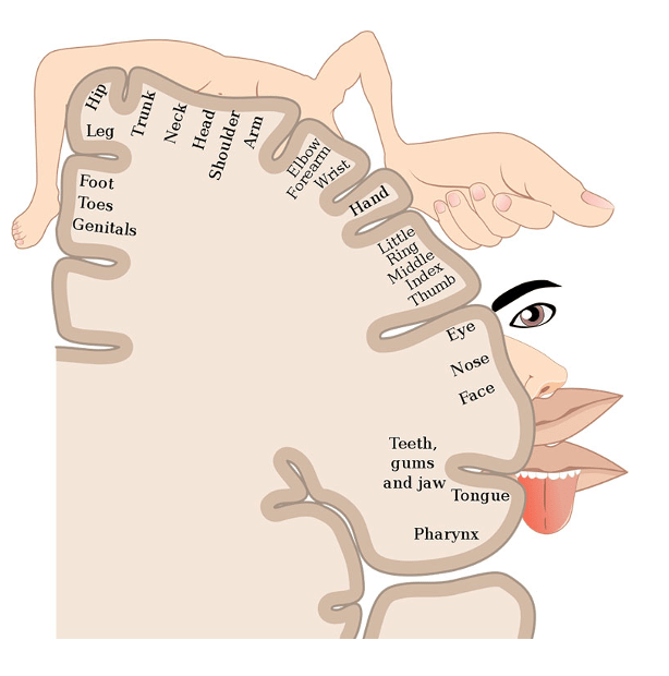
Which body regions occupy the largest proportions of the somatosensory homunculus in the primary somatosensory cortex?
A) Feet and back
B) Torso and arms
C) Face and hands
D) Legs and abdomen
E) Head and neck
Answer: C) Face and hands
Explanation: The face and hands occupy the largest proportions of the somatosensory homunculus because they are highly sensitive areas requiring precise sensory input and motor control. The face is critical for social interactions and conveying emotions, while the hands are essential for fine motor skills and tactile discrimination.
Question 56:
Which of the following best describes the layout of sympathetic and parasympathetic fibres?
A) Short pre-ganglionic fibres and long post-ganglionic fibres for both systems.
B) Long pre-ganglionic fibres and short post-ganglionic fibres for both systems.
C) Short pre-ganglionic fibres and short post-ganglionic fibres for sympathetic, and long pre-ganglionic fibres and long post-ganglionic fibres for parasympathetic.
D) Long pre-ganglionic fibres and short post-ganglionic fibres for sympathetic, and short pre-ganglionic fibres and long post-ganglionic fibres for parasympathetic.
E) Short pre-ganglionic fibres and long post-ganglionic fibres for sympathetic, and long pre-ganglionic fibres and short post-ganglionic fibres for parasympathetic.
Answer: E) Short pre-ganglionic fibres and long post-ganglionic fibres for sympathetic, and long pre-ganglionic fibres and short post-ganglionic fibres for parasympathetic.
Explanation: In the sympathetic nervous system, the preganglionic fibres are relatively short, branching from the spinal cord and synapsing with postganglionic neurons located in sympathetic ganglia near the spinal cord. From there, the postganglionic fibres extend to their target organs. Conversely, in the parasympathetic nervous system, the preganglionic fibres are long, extending from the brainstem or sacral spinal cord to ganglia located near or within the target organs. The postganglionic fibres are short and innervate the effector organs directly.
Question 57:
What type of fibres travel through the grey rami communicans?
A) Pre-ganglionic fibres
B) Sensory fibres
C) Motor fibres
D) Post-ganglionic fibres
E) Sympathetic fibres
Answer: D) Post-ganglionic fibres
Explanation: The grey rami communicans contain post-ganglionic fibres of the sympathetic nervous system. These fibres connect the sympathetic ganglia to the spinal nerves, allowing for the distribution of sympathetic innervation to various parts of the body. They carry nerve impulses away from the autonomic ganglia to effector organs, such as smooth muscle, glands, and blood vessels.
Option A is incorrect because pre-ganglionic fibres travel through the white rami communicans.
Option B is incorrect because sensory fibres do not travel through the grey rami communicans. Sensory fibres travel through dorsal root ganglia.
Option C is incorrect because motor fibres primarily involve the efferent pathways of the somatic nervous system and autonomic nervous system.
Option D is correct because post-ganglionic fibres travel through the grey rami communicans.
Option E is incorrect because sympathetic fibres include both pre-ganglionic and post-ganglionic fibres, but specifically post-ganglionic fibres travel through the grey rami communicans.
Question 58:
What is the main function of Meissner receptors?
A) Detecting changes in temperature
B) Pain detection
C) Sustained touch
D) Discerning fine touch and texture
E) Pressure detection
Answer: D) Discerning fine touch and texture
Explanation: Meissner receptors, also known as tactile corpuscles, are specialised sensory receptors primarily responsible for detecting fine touch and texture. They are particularly abundant in areas such as the fingertips, palms, and lips, contributing to the sense of touch and facilitating tasks such as object manipulation and texture discrimination.
Option A is incorrect because detecting changes in temperature is primarily the function of thermoreceptors aka free nerve endings.
Option B is incorrect because pain detection is primarily mediated by nociceptors.
Option C is incorrect because sustained touch is perceived by Merkel cells, which are located in the skin.
Option E is incorrect because pressure detection is primarily mediated by Pacinian corpuscles and Ruffini endings.

Question 59:
In the central nervous system, where are most third-order neurones found?
A) Somatosensory cortex
B) Spinal Cord
C) Pyramids of medulla oblongata
D) Thalamus
E) Colliculi of midbrain
Answer: D) Thalamus
Explanation: Third-order neurons are typically found in the thalamus, a key relay centre in the brain. These neurons receive sensory information from second-order neurons and transmit it to the cerebral cortex for further processing and interpretation. The thalamus plays a crucial role in sensory perception, motor control, and regulation of consciousness.
Option A is incorrect because the somatosensory cortex receives sensory information from 3rd order neurons but does not house third-order neurons.
Option B is incorrect because the spinal cord primarily contains first-order and second-order neurons involved in transmitting sensory information to higher brain centres.
Option C is incorrect because the pyramids of the medulla oblongata are primarily involved in decussation of neurons.
Option E is incorrect because the colliculi of the midbrain are involved in visual and auditory reflexes.
Question 60:
Which sensations are primarily transmitted through the Dorsal Column-Medial Lemniscus (DCML) pathway?
A) Pressure and unconscious proprioception
B) Pain and temperature
C) Proprioception and crude touch
D) Proprioception, fine touch and vibration
E) Pain, temperature, crude touch and vibration
Answer: D) Proprioception, fine touch, and vibration
Explanation: The Dorsal Column-Medial Lemniscus (DCML) pathway is responsible for transmitting proprioception (awareness of body position), two-point touch discrimination, fine touch sensation, and vibration sense from the body to the brain. This pathway carries sensory information from mechanoreceptors located in the skin, muscles, tendons, and joints to the somatosensory cortex of the brain for processing and perception.
Option A is incorrect because pressure is primarily transmitted through the spinothalamic tract, while unconscious proprioception is a function of the spinocerebellar tract.
Option B is incorrect because pain and temperature sensations are primarily transmitted through the lateral spinothalamic tract.
Option C is incorrect because although conscious proprioception is transmitted through the DCML tract, crude touch is transmitted via the anterior spinothalamic tract.
Option E is incorrect because pain, temperature, and crude touch sensations are primarily transmitted through the spinothalamic tract.
Question 61:
Which specific part of the spinal cord carries signals from the lower part of the body for proprioception, fine touch, and vibration?
A) Cuneocerebellar
B) Lateral spinothalamic tract
C) Ventral posterolateral nucleus
D) Fasciculus cuneatus
E) Fasciculus gracilis
Answer: E) Fasciculus gracilis
Explanation: Signals related to proprioception, fine touch, and vibration from the lower part of the body ascend through the dorsal columns of the spinal cord. Specifically, fasciculus gracilis carries these sensory signals from the lower limbs and lower trunk (below T6 level of the spinal cord) to higher levels of the central nervous system for processing and interpretation.
Option A is incorrect because the cuneocerebellar tract carries proprioceptive information from the upper body to the cerebellum (which is responsible unconscious proprioception & coordination, not fine touch & vibration).
Option B is incorrect because the lateral spinothalamic tract primarily carries pain and temperature sensations & crude touch.
Option C is incorrect because the ventral posterolateral nucleus is a thalamic nucleus that receives sensory input, but it is not a part of the spinal cord.
Option D is incorrect because although fasciculus cuneatus carries impulses regarding proprioception, fine touch, and vibration signals, it carries those impulses from the upper body (above T6).
Question 62:
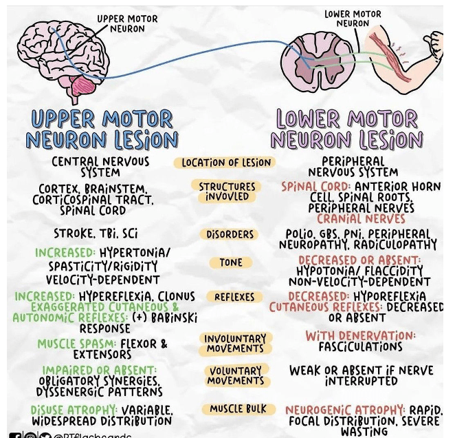
Which set of clinical features is commonly observed in patients with Lower Motor Neuron (LMN) lesions?
A) Increased muscle tone, hyperreflexia, fasciculations
B) Muscle atrophy, spasticity, hyporeflexia, fasciculations
C) Muscle atrophy, flaccidity, weakness, fasciculations, hyporeflexia
D) Flaccidity, clonus, spasticity, hyperreflexia
E) Flaccidity, hyporeflexia, muscle weakness, muscle atrophy, increased tone
Answer: C) Muscle atrophy, flaccidity, weakness, fasciculations, hyporeflexia
Explanation: Lower Motor Neuron (LMN) lesions typically lead to muscle atrophy (due to denervation), flaccidity (loss of muscle tone), weakness (due to loss of motor function), fasciculations (visible twitching of muscle fibres), and hyporeflexia (decreased or absent reflexes). These features reflect the disruption of signals from the spinal cord or brainstem to the muscles, resulting in characteristic clinical findings.
Option A is incorrect because increased muscle tone and hyperreflexia are features commonly associated with Upper Motor Neuron (UMN) lesions. Fasciculations, however, can be seen in LMN lesions.
Option B is incorrect because spasticity is a feature of UMN lesions, not LMN lesions.
Option D is incorrect because spasticity, hyperreflexia and clonus are features of UMN lesions.
Option E is incorrect because increased tone is typically associated with UMN lesions. Weakness is a characteristic of both upper & lower motor neuron lesions.
Question 63:
At which specific location does the corticospinal tract decussate in the central nervous system?
A) Midbrain
B) Cerebral Cortex
C) Olives of the medulla
D) Spinal cord
E) Pyramids of the medulla
Answer: E) Pyramids Medulla
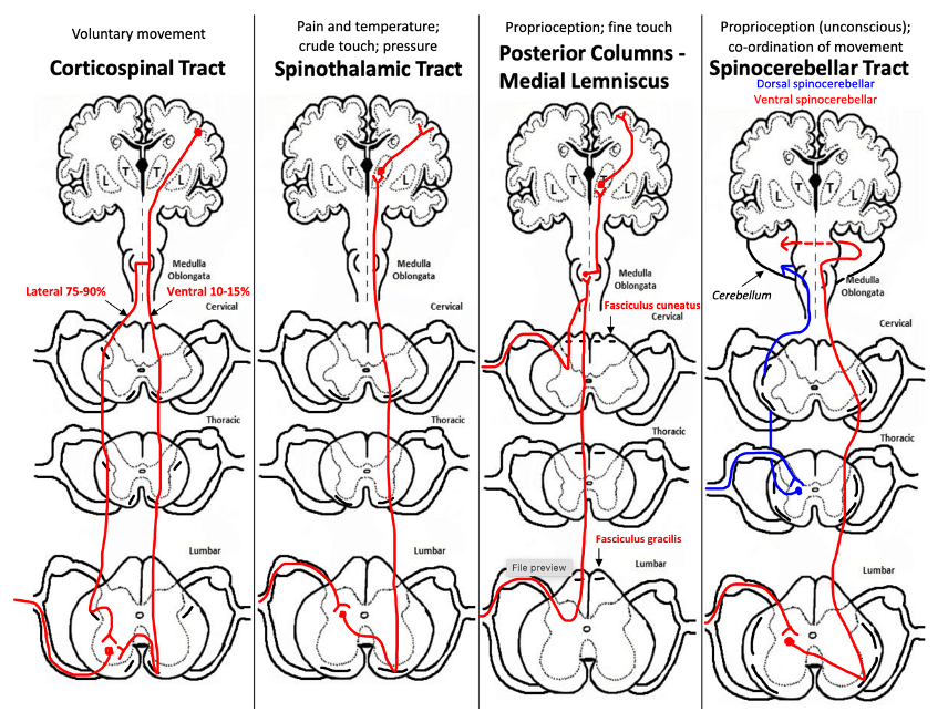
Explanation: The corticospinal tract, also known as the pyramidal tract, decussates (crosses over) in the pyramids of the medulla oblongata of the brainstem. After decussation, the fibres of the tract continue down the spinal cord on the opposite side from where they originated. This crossing allows for contralateral control of voluntary movements: the left hemisphere of the brain controls movements on the right side of the body, and vice versa.
Option A is incorrect because the midbrain is involved in various sensory and motor functions but not in the decussation any tract.
Option B is incorrect because the cerebral cortex is where the corticospinal tract originates (specifically the primary motor cortex).
Option C is incorrect because the olives are structures in the medulla oblongata involved in motor coordination and learning.
Option D is incorrect because the corticospinal tract descends through the spinal cord but does not decussate there.
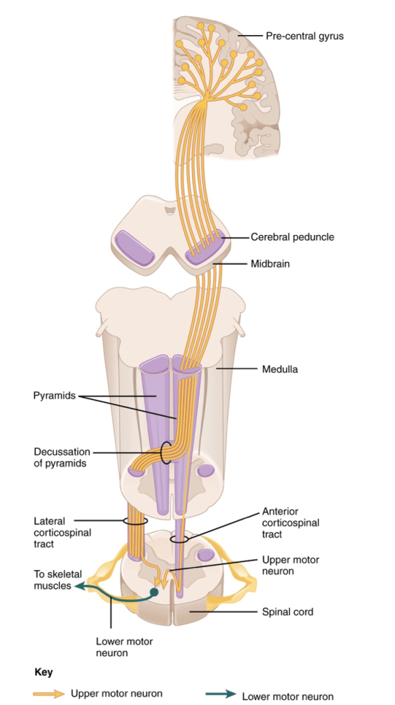
Question 64:
What is the smallest bone in the body?
- Stapes
- Hamate
- Malleus
- Incus
- Navicular
Answer: A) Stapes
Explanation: Stapes is an ossicle of the middle ear & is also the smallest bone in the body. It is connected to incus laterally & the oval window medially.
Sound waves enter the ear and cause the tympanic membrane (eardrum) to vibrate. These vibrations are transmitted to the ossicles (tiny bones of the middle ear) in a sequence.
The vibrations move from the malleus (hammer) to the incus, and then to the stapes. The stapes acts as a bridge between the middle ear and the inner ear.
The stapes’ base is attached to the oval window of the cochlea. When the stapes vibrates, it transmits these vibrations to the fluid-filled cochlea & semi-circular canals, which then converts the mechanical vibrations into electrical signals that are sent to the brain via the vestibulocochlear nerve.
Option B is incorrect because hamate, one of the carpal bones located in the hand that has a hook shape.
Option E is incorrect because navicular a tarsal bone located in the feet.
Options C & D are incorrect because malleus & incus are the other two ossicles located in the middle ear that are also very tiny bones, however are not the smallest bones in the body.
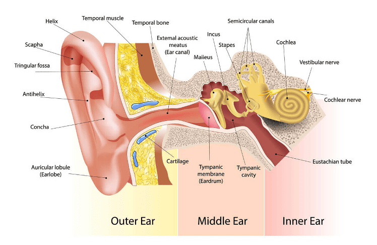
Question 65:
Which structure separates the right & left hemispheres of the brain?
A) Longitudinal sulcus
B) Central sulcus
C) Longitudinal fissure
D) Longitudinal gyrus
E) Central gyrus
Answer: C) Longitudinal fissure
Explanation: The right and left hemispheres of the brain are separated by the longitudinal fissure, which is a deep groove running from the front to the back of the brain. This fissure divides the brain into two distinct hemispheres, allowing each to function independently while maintaining communication through the corpus callosum.
Option B is incorrect because the central sulcus separates the frontal lobe (pre-central gyrus) from the parietal lobe (post-central gyrus).
The lateral fissure separates the frontal & parietal lobes from the temporal lobe.

Question 66:
Where is the primary somatosensory cortex located?
- Parietal lobe
- Post-central gyrus
- Pre-central gyrus
- Temporal lobe
- Frontal lobe
Answer: B) Post-central gyrus
Explanation: The primary somatosensory cortex is located in the post-central gyrus, which is situated in the parietal lobe of the brain. This area is responsible for processing sensory information related to touch, pressure, temperature, and pain from the body which is relayed by the thalamus. The pre-dominant area of the neocortex found in the post-central gyrus, is the 4th layer aka the internal granular layer.
Option A is incorrect because while the primary somatosensory cortex is indeed located in the parietal lobe, the precise location within this lobe is the post-central gyrus therefore option B is a better answer.
Option C is incorrect because the pre-central gyrus, also known as the primary motor cortex, is involved in motor control, and is located in the frontal lobe, not the parietal lobe.
Option D is incorrect because the temporal lobe is involved in auditory processing and memory but does not house the primary somatosensory cortex.
Option E is incorrect because the frontal lobe contains the primary motor cortex and other areas involved in higher cognitive functions such as Broca’s area (language production area) but does not contain the primary somatosensory cortex.
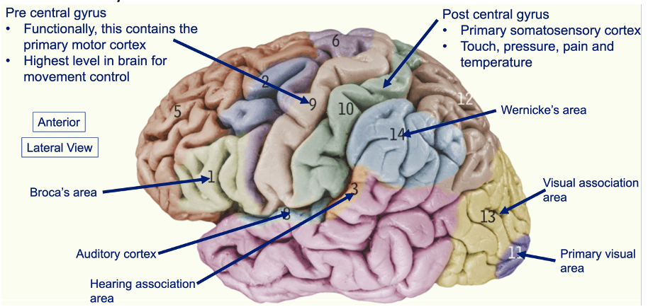
Question 67:
What structure links the endocrine system to the nervous system?
- Thalamus
- Epithalamus
- Subthalamus
- Pituitary gland
- Hypothalamus
Answer: E) Hypothalamus
Explanation: The structure that links the endocrine system to the nervous system is the hypothalamus. The hypothalamus is located in the diencephalon of the brain and plays a crucial role in maintaining homeostasis. It controls the pituitary gland, which is the master gland of the endocrine system. The hypothalamus sends signals to the pituitary gland to regulate hormone release, influencing various bodily functions such as growth, metabolism, and stress responses.
Option A is incorrect because the thalamus is involved in relaying sensory information to the cerebral cortex and does not directly link the endocrine system to the nervous system.
Option B is incorrect because the epithalamus is part of the diencephalon and includes the pineal gland, which regulates circadian rhythms by releasing melatonin.
Option C is incorrect because the subthalamus is involved in motor control and is part of the basal ganglia.
Option D is incorrect because the pituitary gland, while crucial in the endocrine system, is regulated by the hypothalamus and not directly responsible for linking the endocrine system to the nervous system on its own.
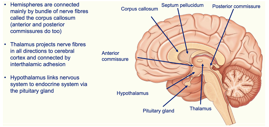
Question 68:
Which structure joins the cerebellum to the medulla oblongata?
- Superior peduncle
- Inferior peduncle
- Meninges
- Calcarine sulcus
- Middle peduncle
Answer: B) Inferior peduncle
Explanation: The structure that joins the cerebellum to the medulla oblongata is the inferior peduncle. The inferior cerebellar peduncle, also known as the restiform body, connects the cerebellum to the medulla oblongata and is involved in relaying sensory information and coordinating motor control.
Option A is incorrect because the superior peduncle connects the cerebellum to the midbrain and is involved in motor coordination and proprioceptive information.
Option C is incorrect because the meninges are protective coverings of the brain and spinal cord. They separate the cerebellum from the occipital lobe.
Option D is incorrect because the calcarine sulcus is a deep groove in the occipital lobe of the brain and is involved in the visual processing area.
Option E is incorrect because the middle peduncle, also known as the middle cerebellar peduncle, connects the cerebellum to the pons and is involved in transmitting information from the cerebral cortex to the cerebellum.
Question 69:
What structures make up the spinocerebellum?
- Vermis & paravermis
- Vermis & primary fissure
- Primary fissure & horizontal fissure
- Vermis, paravermis & cerebellar peduncles
- Horizontal fissure & tonsils
Answer: A) Vermis & paravermis
Explanation: The structures that make up the spinocerebellum are the vermis and the paravermis. The spinocerebellum is a functional division of the cerebellum involved in coordinating and regulating movement. The vermis is the central part of the cerebellum, and the paravermis is the lateral region adjacent to the vermis. Together, these structures integrate sensory input from the spinal cord and are crucial for motor control and coordination.
Question 70:
What is the function of the vestibulocerebellum?
- Unconscious proprioception
- Motor learning
- Planning & coordinating behaviour
- Balance & eye movement
- Regulating movements & conscious proprioception
Answer: D) Balance & eye movement
Explanation: The function of the vestibulocerebellum is balance and eye movement. The vestibulocerebellum, which includes the flocculonodular lobe of the cerebellum, plays a crucial role in maintaining equilibrium and coordinating eye movements to ensure stable vision while the head and body are in motion. It integrates sensory information from the vestibular system (related to balance) and helps coordinate responses to maintain posture and gaze.
Option A is incorrect because unconscious proprioception is primarily managed by the spinocerebellum, which integrates sensory input related to body position and movement without conscious awareness.
Options B & C are incorrect because motor learning, planning & coordinating behaviour is associated with the cerebrocerebellum, which is involved in the planning and refinement of skilled movements through practice & works with the cerebral cortex to plan and execute complex motor activities.
Option E is incorrect because regulating movements are functions of the spinocerebellum and cerebrocerebellum. Conscious proprioception is not associated with the cerebellum.
Question 71:
From which part of the brain/brainstem do cranial nerves III (oculomotor) & IV (trochlear) emerge from?
- Cerebral cortex
- Occipital lobe
- Midbrain
- Medulla oblongata
- Cerebellum
Answer: C) Midbrain
Explanation: Cranial nerves III (oculomotor) and IV (trochlear) emerge from the midbrain. The oculomotor nerve (III) originates from the midbrain’s interpeduncular fossa, while the trochlear nerve (IV) emerges from the dorsal aspect of the midbrain, just below the inferior colliculi. CN V-VIII emerge from the pons & CNs I & II emerge from the olfactory bulb (nose) & eyes respectively.
Option A is incorrect because the cerebral cortex is involved in higher cognitive functions and does not directly give rise to cranial nerves.
Option B is incorrect because the occipital lobe is primarily involved in visual processing but does not give rise to nerves that control ocular muscles & movement.
Option D is incorrect because the medulla oblongata is where cranial nerves IX (glossopharyngeal), X (vagus), XI (accessory), and XII (hypoglossal) emerge.
Option E is incorrect because the cerebellum is responsible for motor control and coordination but does not give rise to any cranial nerves.
Question 72:
Which cranial nerve runs in the cribiform plate?
- Olfactory
- Optic
- Abducens
- Facial
- Trigeminal
Answer: A) Olfactory nerve
Explanation: The cranial nerve that runs in the cribriform plate is the olfactory nerve (I) which is located in the anterior cranial fossa. The olfactory nerve fibres pass through the cribriform plate of the ethmoid bone to reach the olfactory bulbs, where they play a crucial role in the sense of smell.
Option B is incorrect because the optic nerve (II) passes through the optic canal in the sphenoid bone.
Option C is incorrect because the abducens nerve (VI) exits the skull via the superior orbital fissure.
Option D is incorrect because the facial nerve (VII) exits the skull through the stylomastoid foramen and also enters the internal acoustic meatus.
Option E is incorrect because the trigeminal nerve (V) has three branches (ophthalmic, maxillary, and mandibular) that pass through different foramina (supraorbital fissure, foramen rotundum, and foramen ovale respectively).
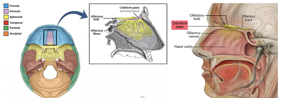

Question 73:
In addition to the vestibulocochlear nerve, what other nerve also passes through the internal acoustic meatus?
- Glossopharyngeal
- Hypoglossal
- Accessory
- Vagus
- Facial
Answer: E) Facial
Explanation: In addition to the vestibulocochlear nerve (VIII), the facial nerve (VII) also passes through the internal acoustic meatus in the posterior cranial fossa. The internal acoustic meatus is a passage in the temporal bone that allows these two cranial nerves to reach their respective destinations. The facial nerve enters the meatus and then travels through the temporal bone to innervate the muscles of facial expression.
Options A , C & D are incorrect because the glossopharyngeal nerve (IX), vagus nerve (X), and accessory nerve (XI) exit the skull through the jugular foramen, not the internal acoustic meatus.
Option B is incorrect because the hypoglossal nerve (XII) passes through the hypoglossal canal, not the internal acoustic meatus.
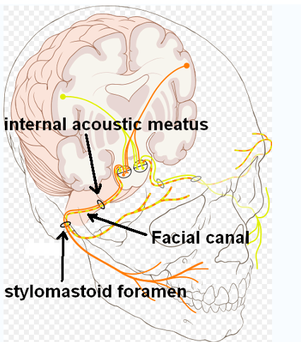
Question 74:
Which of the following is the correct order of the layers of the meninges from deep to superficial?
- Dura, arachnoid, pia
- Pia, arachnoid, dura
- Pia, dura arachnoid, dura
- Arachnoid, pia, dura
- Arachnoid, dura, pia
Answer: B) Pia, arachnoid, dura
Explanation: The pia mater is the innermost layer, closely adhering to the surface of the brain and spinal cord. The arachnoid mater is the middle layer, located between the pia mater and the dura mater. The dura mater is the outermost and most durable layer, providing a tough protective covering.
Pia Mater: Closest to the brain and spinal cord; provides nutrient support and follows the contours of the CNS.
Arachnoid Mater: Middle layer; contains the cerebrospinal fluid and cushions the CNS.
Dura Mater: Outermost layer; provides a tough protective covering.

Question 75:
Which of the following ventricles of the brain secrete the greatest volume of cerebrospinal fluid?
- 4th ventricle
- Cerebral aqueduct (Sylvian aqueduct)
- 3rd ventricle
- Interventricular foramen (Foramen of Monro)
- Lateral ventricles
Answer: E) Lateral ventricles
Explanation: The ventricles of the brain that secrete the greatest volume of cerebrospinal fluid (CSF) are the lateral ventricles. These ventricles, located in each cerebral hemisphere, produce the majority of the brain’s CSF via the choroid plexus found in them. The CSF then flows from the lateral ventricles to the 3rd ventricle via the interventricular foramen. From the 3rd ventricle, CSF flows into the 4th ventricle via the cerebral aqueduct. CSF enters from the subarachnoid space via the median aperture medially (Foramen of Magendie) & lateral aperture laterally (Foramen of Luschka). CSF leaves the subarachnoid space & enters the bloodstream via the superior sagittal sinus.
Options A & C are incorrect because the 3rd & 4th ventricle produces some CSF but not as much as the lateral ventricles.
Option B is incorrect because the cerebral aqueduct (Sylvian aqueduct) connects the 3rd and 4th ventricles and does not produce CSF.
Option D is incorrect because the interventricular foramen is a channel that connects the lateral ventricles to the 3rd ventricle and does not produce CSF.
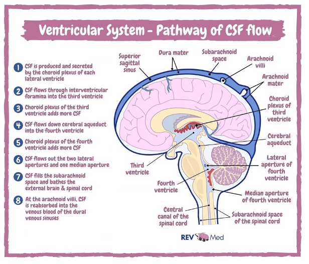
Question 76:
Which 2 arteries provide the majority of the blood supply to the brain?
- External carotid & vertebral arteries
- Internal carotid & vertebral arteries
- Common carotid & vertebral arteries
- Internal carotid & subclavian arteries
- Internal carotid & brachiocephalic arteries
Answer: B) Internal carotid & vertebral arteries
Explanation: The two arteries that provide the majority of the blood supply to the brain are the internal carotid and vertebral arteries. The internal carotid arteries supply the anterior circulation of the brain, including the frontal, parietal, and parts of the temporal lobes, while the vertebral arteries supply the posterior circulation, including the brainstem, cerebellum, and occipital lobes. These arteries contribute to the formation of the circle of Willis, which is crucial for maintaining cerebral blood flow.
Option A is incorrect because the external carotid artery primarily supplies blood to the face and scalp.
Option C is incorrect because the common carotid artery bifurcates into the internal and external carotid arteries, but on its own, it does not supply the brain directly.
Option D is incorrect because the subclavian artery supplies the upper limbs and parts of the thorax.
Option E is incorrect because the brachiocephalic artery supplies blood to the right side of the head and right arm but does not directly supply the brain.
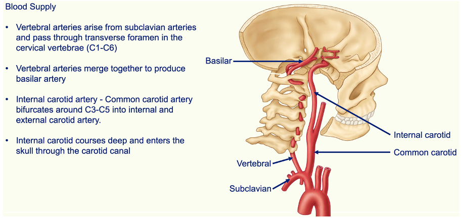

Question 77:
What is the predominant venous drainage of the brain?
- Internal jugular vein
- Confluence of sinus
- Superior sagittal sinus
- Great cerebellar vein
- Cavernous sinus
Answer: A) Internal jugular vein
Explanation: The venous drainage of the brain primarily involves the internal jugular vein. The internal jugular vein receives blood from the dural venous sinuses, which drain the brain. Blood from these sinuses flows into the internal jugular vein, which then carries it back to the heart.
Option B is incorrect because while the confluence of sinuses is a key junction where several major dural sinuses meet, including the superior sagittal sinus, it is not the final pathway for venous drainage.
Option C is incorrect because the superior sagittal sinus is one of the major dural venous sinuses that collects blood from the brain, but it is not the final vessel for venous drainage.
Option D is incorrect because the great cerebellar vein primarily drains the cerebellum.
Option E is incorrect because the cavernous sinus is another important dural venous sinus that drains blood from the brain, but it is not the final vessel responsible for venous drainage.

Question 78:
At which vertebral level does the adult spinal cord terminate?
- T12 – L1
- L1 – L2
- L2 – L3
- L3 – L4
- L4 – L5
Answer: B) L1 – L2
Explanation: The adult spinal cord typically terminates at the level of L1 – L2. This is where the conus medullaris, the tapered end of the spinal cord, is located. Beyond this point, the spinal nerves continue as the cauda equina.

Question 79:
Which of the following covers the posterior & lateral 5/6th of the fibrous layer of the eye?
- Cornea
- Sclera
- Choroid
- Macula lutea
- Optic disc
Answer: B) Sclera
Explanation: The structure that covers the posterior and lateral 5/6th of the fibrous layer of the eye is the sclera. The sclera is the white, opaque part of the eye that provides structural support and protection. It forms the bulk of the fibrous layer, which is the outermost layer of the eye.
Option A is incorrect because the cornea covers the anterior 1/6th of the eye’s surface and is responsible for focusing light, but it does not cover the posterior and lateral portions.
Option C is incorrect because the choroid is a vascular layer of the eye (2nd layer) located between the retina and the sclera, providing nourishment to the retina, but it is not part of the fibrous layer.
Option D is incorrect because the macula lutea is an area of the retina responsible for central vision and does not cover any part of the fibrous layer.
Option E is incorrect because the optic disc is the point where the optic nerve exits the eye.
Question 80:
Which of the following make up the 3 layers of the eye?
- Fibrous, vascular, retina
- Fibrous, ciliary, retina
- Fibrous, muscular, retina
- Fibrous, muscular, vascular
- Fibrous, ciliary, vascular
Answer: A) Fibrous, vascular, retina
Explanation: The three layers of the eye are the fibrous layer, the vascular layer, and the retina. The fibrous layer consists of the sclera and cornea, providing structural support and protection to the eye. The vascular layer, also known as the uvea, includes the choroid, ciliary body, and iris. It is responsible for supplying blood to the eye and controlling light entry. The retina is the innermost layer of the eye where photoreceptors are located, enabling the conversion of light into neural signals that are sent to the brain.
The ciliary layer is part of the vascular layer, not a separate layer on its own.
The muscular layer is not one of the primary layers of the eye.
Question 81:
Which of the following eye muscles does the sympathetic nervous system innervate?
- Sphincter pupillae
- Superior oblique
- Inferior oblique
- Inferior rectus
- Dilator pupillae
Answer: E) Dilator pupillae
Explanation: The muscle innervated by the sympathetic nervous system is the dilator pupillae which is located in the iris (vascular part of the eye). This muscle is responsible for dilating the pupil in response to low light or sympathetic stimulation, such as during the “fight or flight” response.
Option A is incorrect because the sphincter pupillae is controlled by the parasympathetic nervous system and is responsible for constricting the pupil.
Option B is incorrect because the superior oblique muscle is innervated by the trochlear nerve (cranial nerve IV).
Option C is incorrect because the inferior oblique muscle is innervated by the oculomotor nerve (cranial nerve III).
Option D is incorrect because the inferior rectus muscle is also innervated by the oculomotor nerve (cranial nerve III).

Question 82:
Which section of the eye is the thinnest & is where the majority of cones (colour photoreceptors) are located?
- Ora Serrata
- Optic disc
- Macula lutea
- Fovea
- Retina
Answer: D) Fovea
Explanation: The thinnest section of the eye where the majority of cones (colour photoreceptors) are located, is the fovea. The fovea is a small, central pit in the macula lutea of the retina and is specialised for sharp central vision (high visual acuity) and colour perception due to its high density of cone cells.
Option A is incorrect because the ora serrata is the boundary between the retina and the ciliary body. It is the first region of the retina that contains photoreceptors (rods) & can detect light but does not have the highest concentration of cones.
Option B is incorrect because the optic disc is where the optic nerve exits the eye and lacks photoreceptors; it is known as the blind spot.
Option C is incorrect because the macula lutea is a region of the retina surrounding the fovea that contains a high concentration of cones, but the fovea itself is the thinnest and has the highest density of these photoreceptors.
Option E is incorrect because the retina is the layer of the eye that contains both rods and cones, but it is not the thinnest section nor the specific area with the highest concentration of cones.
Question 83:
Which photoreceptors connect to the optic nerve?
- Rods & cones
- Horizontal cell
- Bipolar cells
- Amacrine cells
- Ganglion cells
Answer: E) Ganglion cells
Explanation: The photoreceptors that connect to the optic nerve are the ganglion cells. Ganglion cells are the final output neurons of the retina, whose axons form the optic nerve, carrying visual information from the retina to the brain.
Option A is incorrect because rods and cones are the photoreceptors responsible for detecting light and colour, but they do not directly connect to the optic nerve. Instead, they synapse with bipolar cells.
Options B & D are incorrect because horizontal cells and amacrine cells are interneurons that modulate the signals between photoreceptors, bipolar cells, and ganglion cells, but they do not connect directly to the optic nerve.
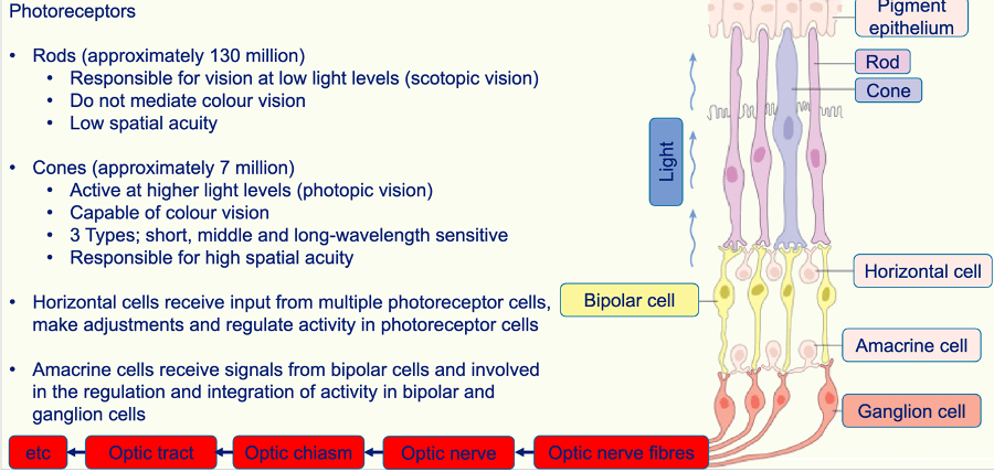
Question 84:
Which of the following muscles elevates, adducts & medially rotates the eye?
- Superior oblique
- Superior rectus
- Inferior oblique
- Inferior rectus
- Levator palpabrae superioris
Answer: B) Superior rectus
Explanation: The superior rectus muscle is one of the extraocular muscles and is responsible for moving the eye upwards (elevation), towards the nose (adduction), and medially rotating it.
Option A is incorrect because he superior oblique muscle primarily depresses, abducts, and medially rotates the eye.
Option C is incorrect because the inferior oblique muscle elevates, abducts, and laterally rotates the eye.
Option D is incorrect because the inferior rectus muscle primarily depresses, adducts, and laterally rotates the eye.
Option E is incorrect because the levator palpebrae superioris muscle primarily elevates the upper eyelid, not the eye itself, and does not contribute to adduction or medial rotation.
General rule:
- Recti muscles adduct
- Oblique muscles abduct
- Superiors intort
- Inferiors extort
- Recti muscles move in the same direction (superior rectus elevates & inferior rectus depresses)
- Oblique muscles move in the opposite direction (superior oblique depress & inferior oblique elevates).
- Superior muscles medially rotate (superior oblique & superior rectus medially rotate)
- Inferior muscles laterally rotate (inferior oblique & inferior rectus laterally rotate)
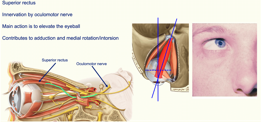
Question 85:
Which part of the ear is the region where sound waves enter?
- External acoustic meatus
- Lobule
- Concha
- Tragus
- Helix
Answer: A) External acoustic meatus
Explanation: The part of the ear where sound waves enter is the external acoustic meatus. The external acoustic meatus is the ear canal that directs sound waves from the external environment to the tympanic membrane (eardrum). The tympanic membrane transmits vibration to ossicles of the middle ear which then then carry those vibrations to the labyrinth of the inner ear.
Option B is incorrect because the lobule is the soft, fleshy part of the ear known as the earlobe where kids have their ears pierces. It does not play a role in directing sound waves.
Option C is incorrect because the concha is the bowl-shaped part of the outer ear that helps funnel sound waves into the external acoustic meatus but is not where sound waves directly enter.
Option D is incorrect because the tragus is a small, protruding structure at the front of the ear canal, which also helps to direct sound but is not the main entry point for sound waves.
Option E is incorrect because the helix is the outer rim of the ear, which provides structure but does not directly channel sound waves into the ear canal.
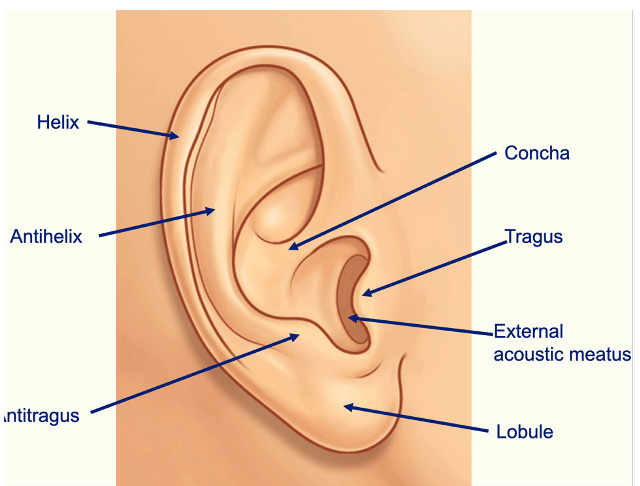
Question 86:
Which of the following fluids fill the bony labyrinth of the inner ear?
- Endosteum
- Endolymph
- Perilymph
- Periosteum
- Epiosteum
Answer: C) Perilymph
Explanation: The bony labyrinth, which is the outer bony structure of the inner ear, contains this fluid that surrounds the membranous labyrinth. The perilymph acts as a cushion and helps to transmit sound vibrations from the oval window to the fluid inside the cochlea.
Option A is incorrect because endosteum lines the inner surface of bones but does not fill the bony labyrinth.
Option B is incorrect because endolymph is the fluid found within the membranous labyrinth, not the bony labyrinth.
Option D is incorrect because the periosteum is a connective tissue layer covering the outer surface of bones, including the bony labyrinth, but does not fill it.
Option E is incorrect because the epiosteum is not an anatomical term.
Periosteum lines the bony labyrinth & perilymph fills it.
Question 87:
What structures make up membranous labyrinth of the inner ear?
- Vestibule, semicircular canals & cochlea
- Semicircular ducts, utricle, saccule & cochlear ducts
- Vestibule, utricle & saccule
- Semicircular canals, utricle & saccule
- Semicircular ducts, utricle, saccule, cochlear ducts & vestibule
Answer: B) Semicircular ducts, utricle, saccule & cochlear ducts
Explanation: The membranous labyrinth of the inner ear includes the semicircular ducts, utricle, saccule, and cochlear duct. This system is a network of fluid-filled tubes and sacs within the bony labyrinth, which is crucial for both balance and hearing.
Options A, C, D & E are incorrect because the vestibule and semicircular canals are part of the bony labyrinth.
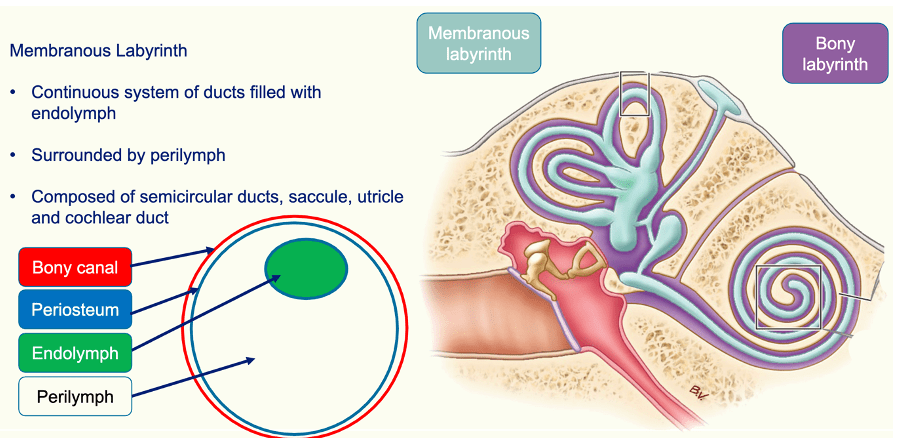
Question 88:
Which of the following triggers the release of neurotransmitters in the vestibulocochlear nerve when vibration is detected?
- Enolymph
- Basilar membrane
- Hair cells
- Sterocilia
- Organ of corti
Answer: C) Hair cells
Explanation: Hair cells in the cochlea are responsible for detecting vibrations and converting them into electrical signals. When these hair cells are stimulated by vibrations, they trigger the release of neurotransmitters, which then transmit the signal through the vestibulocochlear nerve to the brain.
The oval window is moved by the movement of the ossicles. This creates waves that travel through the fluid. These waves cause movement to a structure called the basilar membrane. Different portions of the basilar membrane respond to different frequencies of sound. Waves pass down the basilar membrane & reach their peak at the part of the membrane
that responds to the original stimulus. Movement in the basilar membrane is converted into
an electrical stimulus at the Organ of Corti. At the Organ of Corti stereocilia of hair cells release neurotransmitters to propagate the auditory signal to the vestibulocochlear nerve.
Option A is incorrect because the enolymph is not a structure but a fluid in the inner ear. It helps to transmit sound waves but does not directly trigger neurotransmitter release.
Option B is incorrect because the basilar membrane supports the hair cells but does not itself trigger neurotransmitter release.
Option D is incorrect because sterocilia are the tiny hair-like structures on the hair cells that detect vibrations but do not directly release neurotransmitters.
Option E is incorrect because the organ of Corti houses the hair cells and other structures necessary for hearing, but it is the hair cells specifically that are responsible for neurotransmitter release.

Question 89:
To which region of the thalamus do fibres of the optic nerve project to?
- Lateral geniculate body
- Medial geniculate body
- Betz cells
- Optic radiation
- Lateral lemniscus
Answer: A) Lateral geniculate body
Explanation: The fibres of the optic nerve project to the lateral geniculate body of the thalamus. This region is responsible for processing visual information before it is sent to the primary visual cortex in the occipital lobe.
Option B is incorrect because the medial geniculate body is involved in processing auditory fibres from the vestibulocochlear nerve.
Option C is incorrect because Betz cells are large pyramidal cells located in the motor cortex, not involved in the visual pathway.
Option D is incorrect because the optic radiation is a pathway that carries visual information from the lateral geniculate body to the primary visual cortex, but it is not where the optic nerve fibers initially project.
Option E is incorrect because the lateral lemniscus is part of the auditory pathway and is not related to the visual pathway.
Question 90:
In which of the following structures is endolymph found?
- Scala tympani
- Scala vestibuli
- Scala media
- Tunica media
- Basilar membrane
Answer: C) Scala media
Explanation: Endolymph is found in the scala media, also known as the cochlear duct. The scala media is part of the membranous labyrinth of the inner ear and contains endolymph, which plays a crucial role in the transduction of sound vibrations into neural signals.
Options A & B are incorrect because the scala tympani and scala vestibuli are filled with perilymph, not endolymph.
Option D is incorrect because the tunica media is a layer of blood vessels.
Option E is incorrect because the basilar membrane is a structure within the cochlea but does not contain endolymph.
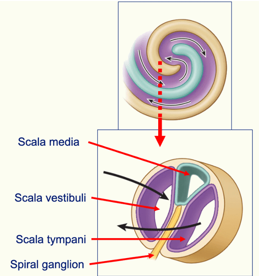
Question 91:
What is the structure of sensory neuron?
A) Bipolar neuron
B) Pseudounipolar neuron
C) Interneuron
D) Multipolar neuron
E) Dendritic neuron
Answer: B) Pseudounipolar neuron
Explanation: Sensory neurons are typically pseudounipolar neurons. In these neurons, a single axon branches into two distinct processes: one extending to the periphery to receive sensory information and the other extending to the central nervous system to transmit that information.
Option A is incorrect because bipolar neurons are commonly found in sensory organs such as the retina and olfactory system but are not the primary structure of sensory neurons throughout the nervous system.
Option C is incorrect because interneurons are primarily involved in relaying information between neurons within the central nervous system and are usually multipolar.
Option D is incorrect because multipolar neurons are the most common type of neurons in the central nervous system and are involved in motor and interneuronal functions rather than sensory functions.
Option E is incorrect because dendritic neurons are not a recognised classification for a sensory neuron.
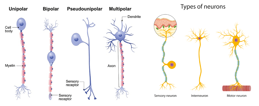
Question 92:
Which of the following structures lines the olfactory nerve (CN I)?
- Olfactory ensheathing cell
- Schwann cell
- Oligodendrocytes
- Astrocytes
- Satellite glial cell
Answer: A) Olfactory ensheathing cell
Explanation: The olfactory nerve (CN I) is lined by olfactory ensheathing cells (OECs). These unique glial cells support the growth and guidance of olfactory neurons from the nasal epithelium to the olfactory bulb and can assist in the regeneration of olfactory nerve fibers.
Option B is incorrect because schwann cells are responsible for myelinating peripheral nerves but are not involved in lining the olfactory nerve.
Option C is incorrect because oligodendrocytes myelinate neurons in the central nervous system (CNS), but the olfactory nerve is lined by OECs rather than oligodendrocytes.
Option D is incorrect because astrocytes provide support and nutrition to neurons in the CNS and maintain the blood-brain barrier.
Option E is incorrect because satellite glial cells support neurons in the peripheral ganglia.
Question :93:
Which of the following is the only unmyelinated nerve (out of the 12 cranial nerves)?
- Hypoglossal (CN XII)
- Accessory (CN XI)
- Vagus (CN X)
- Optic (CN II)
- Olfactory (CN I)
Answer: E) Olfactory (CN I)
Explanation: The only unmyelinated nerve out of the 12 cranial nerves is the olfactory nerve (CN I). This nerve is responsible for the sense of smell and consists of unmyelinated sensory fibres that originate in the olfactory epithelium and project to the olfactory bulb. Despite it being unmyelinated, it is coated with olfactory ensheathing cells which provides the olfactory nerve with protection & support.
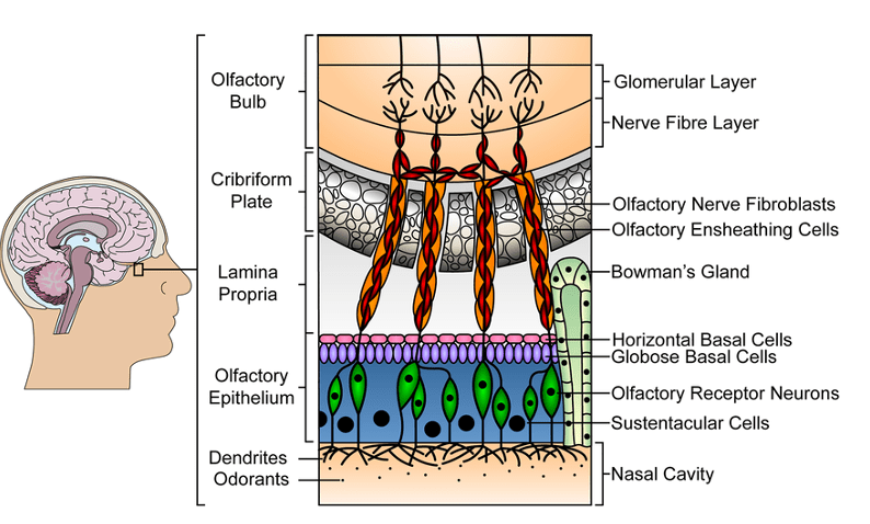
Question 94:
Damage to the endoneurium & perineurium of a neuron describes which of the following conditions?
- Grade I neuropraxia
- Grade II axonotmesis
- Grade III neurotmesis
- Grade IV neurotmesis
- Grade V neurotmesis
Answer: D) Grade IV neurotmesis
Explanation: Damage to the endoneurium and perineurium of a neuron describes Grade IV neurotmesis. This type of nerve injury is severe, involving the disruption of the axon, myelin sheath, endoneurium, and perineurium, with only the epineurium remaining intact. This results in a loss of nerve continuity and typically requires surgical intervention for recovery.
Option A is incorrect because grade I neuropraxia is the mildest form of nerve injury, characterised by a temporary conduction block with no structural damage to the nerve fibres or connective tissue (e.g. resting on your arm).
Option B is incorrect because grade II axonotmesis involves damage to the axon and myelin sheath, but the endoneurium, perineurium, and epineurium remain intact.
Option C is incorrect because grade III neurotmesis involves damage to the axon, myelin sheath, and endoneurium, with the perineurium and epineurium remaining intact.
Option E is incorrect because grade V neurotmesis is the most severe form, with complete transection of the nerve (endoneurium, perineurium & epineurium), resulting in a loss of continuity of the entire nerve trunk and typically requiring surgical repair.

Question 95:
Which of the following layers of the neocortex is predominantly found in the pre-central gyrus?
- Molecular
- External granular
- External pyramidal
- Internal granular
- Internal pyramidal
Answer: E) Internal pyramidal
Explanation: The cerebral cortex is composed of six layers of different types of cells, collectively known as the neocortex. The thickness and abundance of these layers vary between cortical areas of the brain. The predominant layer found in the neocortex of the pre-central gyrus is the internal pyramidal layer (layer V). The pre-central gyrus contains the primary motor cortex, which is responsible for voluntary motor function. This layer is characterized by large pyramidal cells (Betz cells) that project to the spinal cord and brainstem, playing a crucial role in motor control.
Option A is incorrect because the molecular layer (layer I) contains few neurons and consists mainly of horizontal fibres.
Option B is incorrect because the external granular layer (layer II) contains small granular cells and receives input from other cortical areas.
Option C is incorrect because the external pyramidal layer (layer III) contains pyramidal cells that project to other cortical areas.
Option D is incorrect because the internal granular layer (layer IV) contains densely packed stellate cells and is the primary recipient of thalamic input, particularly in somatosensory cortex (post-central gyrus).
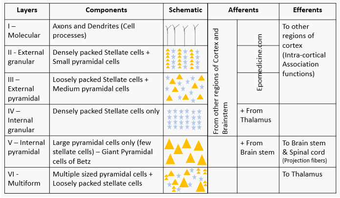
Question 96:
Through which region of a neuron do sodium & potassium ions diffuse into the axon via voltage gated channels?
- Axon hillock
- Axon terminal
- Nodes of Ranvier
- Synaptic cleft
- Pre-synaptic membrane
Answer: C) Nodes of Ranvier
Explanation: Sodium and potassium ions diffuse into the axon via voltage-gated channels primarily at the Nodes of Ranvier. These are small gaps in the myelin sheath where the axon membrane is exposed and there is no myelin in that area. The voltage-gated sodium channels open in response to an action potential, allowing sodium ions to rush in, followed by the opening of potassium channels to allow potassium ions to flow out. This process is crucial for the propagation of action potentials along myelinated axons, enabling rapid and efficient signal transmission.
Option A is incorrect because the axon hillock is the initial segment of the axon where action potentials are generated but does not contain the primary voltage-gated channels for sodium and potassium diffusion along the axon.
Option B is incorrect because the axon terminal is the end part of the axon that releases neurotransmitters into the synaptic cleft. It contains calcium voltage gated ion channels that are necessary in neurotransmitter release, but not sodium or potassium.
Option D is incorrect because the synaptic cleft is the gap between pre & post synaptic neurons at a synapse and is involved in neurotransmitter diffusion, not ion diffusion along the axon.
Option E is incorrect because the pre-synaptic membrane is part of the axon terminal involved in releasing neurotransmitters but is not the primary site for voltage-gated sodium and potassium ion channels.
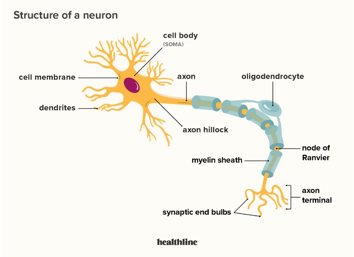
Question 97:
Which of the following is an inhibitory neurotransmitter?
- Aspartate
- Nitric oxide
- Glutamate
- Dopamine
- Acetylcholine
Answer: D) Dopamine
Explanation: Dopamine plays a key role in regulating mood, reward, and motor control. Dopamine has complex effects, as it can act as both an excitatory and inhibitory neurotransmitter depending on the type of receptor it binds to but is generally an inhibitory neurotransmitter.
Option A is incorrect because aspartate is an excitatory neurotransmitter that works in a manner similar to glutamate.
Option B is incorrect because nitric oxide is a gaseous neurotransmitter that primarily functions as a signalling molecule rather than a classic neurotransmitter with direct excitatory or inhibitory properties.
Option C is incorrect because glutamate is the main excitatory neurotransmitter in the brain, involved in most of the brain’s excitatory signalling and cognitive functions.
Option E is incorrect because acetylcholine can act as both an excitatory and inhibitory neurotransmitter, depending on the type of receptor it binds to. For example, it is excitatory at neuromuscular junctions but can be inhibitory in the heart. Therefore, dopamine is a better answer.

Question 98:
Which of the following is a neurotransmitter derived from amino acids?
- Glutamate
- Endorphins
- Substance P
- Adenosine
- Noradrenaline
Answer: A) Glutamate
Explanation: Glutamate is a neurotransmitter derived from amino acids. It is the primary excitatory neurotransmitter in the brain and plays a crucial role in synaptic transmission and plasticity.
Option B is incorrect because endorphins are peptides that act as neurotransmitters, produced from the larger precursor protein pro-opiomelanocortin (POMC) aka neuropeptides.
Option C is incorrect because substance P is a non-opioid peptide neurotransmitter involved in pain perception and inflammatory responses.
Option D is incorrect because adenosine is a purine nucleoside neurotransmitter derived from ATP.
Option E is incorrect because noradrenaline (norepinephrine) is a neurotransmitter derived from the amino acid tyrosine, but it is a catecholamine, which is a class of neurotransmitters distinct from simple amino acid neurotransmitters like glutamate.
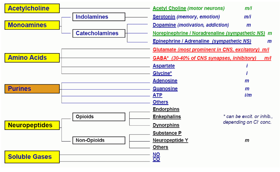
Question 99:
Which of the following upregulates the AMPA receptor?
A) Magnesium ions
B) NMDA receptor
C) Chloride ion receptor
D) Muscarinic receptor
E) Adrenergic receptor
Answer: B) NMDA receptor
Explanation: The NMDA receptor, when activated, facilitates the process of synaptic plasticity and enhances the response of AMPA receptors, which are critical for fast excitatory synaptic transmission and long-term potentiation.
The NMDA receptor is activated through the following steps:
- Ligand Binding: Glutamate and a co-agonist (glycine or D-serine) bind to the NMDA receptor.
- Membrane Depolarisation: The postsynaptic membrane must be depolarised to remove the magnesium ion blockade from the NMDA receptor channel.
- Ion Flow: Once the magnesium block is removed, calcium (Ca²⁺) and sodium (Na⁺) ions flow through the receptor, initiating intracellular signalling and synaptic plasticity.
This activation process is essential for learning and memory by modifying synaptic strength.
Option A is incorrect because magnesium ions act as blockers for NMDA receptors, preventing their activation unless removed by depolarisation.
Option C is incorrect because chloride ion receptors, such as GABA receptors, are generally inhibitory and do not have a direct role in upregulating AMPA receptors.
Option D is incorrect because muscarinic receptors are a type of acetylcholine receptor that can modulate various physiological functions but do not directly upregulate AMPA receptors.
Option E is incorrect because adrenergic receptors are involved in the response to adrenaline and noradrenaline, affecting various physiological processes, but they do not directly upregulate AMPA receptors.
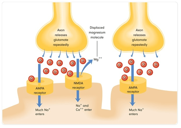
Question 100:
Which of the following fibres are the largest, fastest & most myelinated?
- A-alpha fibre
- A-gamma fibre
- A-delta fibre
- B fibre
- C fibre
Answer: A) A-alpha fibre
Explanation: A-alpha fibres are characterised by their large diameter and thick myelin sheath, which allow them to conduct nerve impulses at high speeds. They are primarily responsible for motor functions and proprioception.
Option B is incorrect because A-gamma fibres are smaller than A-alpha fibres and are involved in motor control of muscle spindles.
Option C is incorrect because A-delta fibres are smaller and less myelinated than A-alpha fibres, involved in transmitting fast, sharp pain and temperature sensations.
Option D is incorrect because B fibres are intermediate in size and myelination, typically associated with autonomic functions.
Option E is incorrect because C fibres are the smallest and unmyelinated, responsible for transmitting slow, dull pain and temperature sensations.

Question 101:
Which of the following contains pre-ganglionic neurons of the parasympathetic nervous system?
- CN III, IV, V & VI
- CN IX, X & XI
- CN III, VII, IX & X
- CN III, VII, IX, X & XI
- CN III, IV, VI, IX & X
Answer: C) CN III, VII, IX & X
Explanation: The parasympathetic nervous system’s pre-ganglionic neurons are primarily found in the following cranial nerves:
CN III (Oculomotor nerve): Controls constriction of the pupil and accommodation of the lens.
CN VII (Facial nerve): Innervates salivary glands (submandibular and sublingual) and lacrimal glands.
CN IX (Glossopharyngeal nerve): Innervates the parotid gland.
CN X (Vagus nerve): Extends its parasympathetic effects to the heart, lungs, and digestive tract.
These cranial nerves contain pre-ganglionic parasympathetic fibres that synapse in ganglia close to or within the target organs.
The parasympathetic functions of these specific cranial nerves help regulate and maintain various body processes such as digestion, heart rate, and glandular secretions, contributing to the body’s rest-and-digest responses.
Question 102:
Which of the following receptors is responsible for detecting sustained touch & pressure?
- Pacinian corpuscle
- Meissner’s corpuscle
- Merkel’s discs
- Ruffini receptors
- Free nerve endings
Answer: C) Merkel’s discs
Explanation: Merkel’s discs are responsible for detecting sustained touch and pressure. They are slowly adapting mechanoreceptors located in the skin and are important for detecting steady pressure and texture.
Option A is incorrect because Pacinian corpuscles detect deep pressure and rapid vibrations (200-300 Hz) and are rapidly adapting, which means they respond quickly to changes rather than sustained pressure.
Option B is incorrect because Meissner’s corpuscles detect light touch and texture changes with rapid adaptation, responding to stimuli around 50 Hz.
Option D is incorrect because Ruffini receptors are slow-adapting mechanoreceptors that respond to deep skin tension, stretch, and joint angle changes. They help modulate grip and provide information about deep pressure and skin stretch.
Option E is incorrect because free nerve endings are not specialized for sustained touch and pressure but respond to a range of stimuli including pain, temperature, and mechanical deformation.
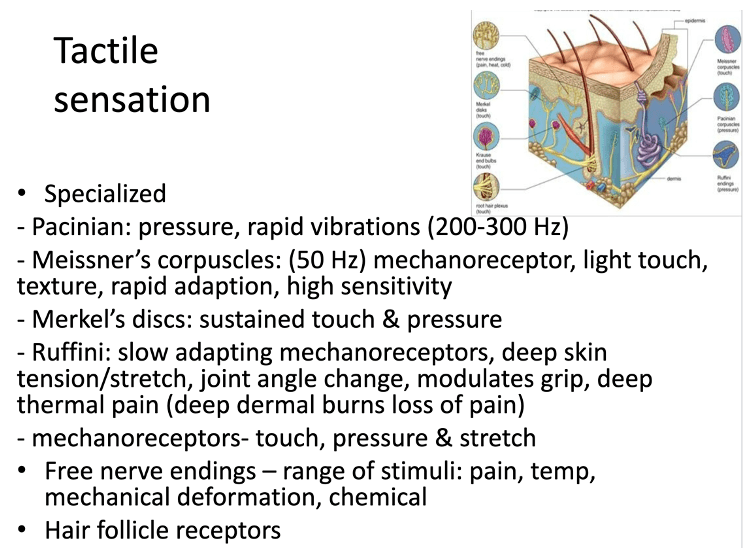
Question 103:
Lesions in the spinal cord causes damage to which of the following tracts resulting in: ipsilateral loss of vibration & proprioception and contralateral loss of pain of temperature known as Brown-Sequard Syndrome?
- Corticospinal tract & spinothalamic tract
- Spinothalamic tract & dorsal column medial lemniscus tract
- Dorsal column medial lemniscus tract & corticospinal tract
- Dorsal column medial lemniscus tract & cerebrocerebellar tract
- Cerebrocerebellar tract & spinothalamic tract
Answer: B) Spinothalamic tract & dorsal column medial lemniscus tract
Explanation: Lesions in the spinal cord causing Brown-Sequard Syndrome typically involve damage to both the dorsal column medial lemniscus (DCML) tract and the spinothalamic tract. The DCML tract carries sensations of vibration and proprioception, so damage to this tract results in ipsilateral (same side) loss of these sensations. The spinothalamic tract is responsible for transmitting pain and temperature sensations; thus, damage to this tract causes contralateral (opposite side) loss of pain and temperature sensations.

Question 104:
Which of the following dermatomes is the referred region of pain involved in appendicitis?
- T6
- T8
- T10
- T12
- L2
Answer: C) T10
Explanation: The referred pain from appendicitis is typically felt in the T10 dermatome. This is because the appendix is innervated by the lower thoracic nerves, primarily around the T10 level. Referred pain occurs when the brain perceives pain in a region of the skin that shares the same spinal nerve segment as the internal organ involved.
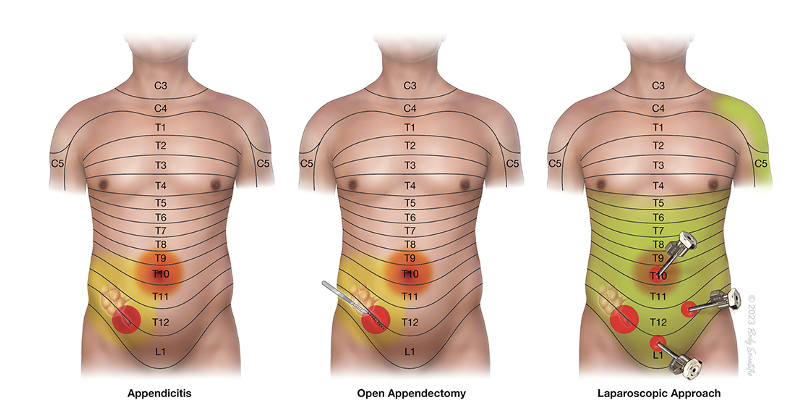
Question 105:
How many synapses are there in the brachial plexus?
- 0
- 1,000
- 10,000
- 100,000
- 1,000,000
Answer: A) 0 synapses
Explanation: The brachial plexus is a network of nerves that originate from the spinal cord in the neck and supply the arm. It is composed of roots, trunks, divisions, cords, and branches. The brachial plexus itself is made up of nerve fibres, which are axons of neurons. Synapses occur between neurons. In the brachial plexus, the nerve fibres are mostly axons, and synapses typically occur in ganglia or at neuromuscular junctions, not within the plexus itself. Therefore, there are no synapses within the brachial plexus itself or in any other plexus in the body.
Question 106:
Which of the following cells inhibits motor neurons whose target is the antagonist muscle in the reflex inhibition of antagonist muscle reflex?
- Betz cells
- Pyramidal cells
- Otolith organs
- Renshaw cell
- Purkinje cell
Answer: D) Renshaw cell
Explanation: When an Ia afferent neuron detects stretch in muscle spindles, it triggers the activation of an alpha motor neuron to cause contraction of the stretched muscle (agonist e.g. biceps). It also stimulates Renshaw cells to inhibit the antagonist muscle (e.g. triceps). Renshaw cells are a type of inhibitory interneuron found in the spinal cord. They receive input from alpha motor neurons and provide feedback inhibition to those same motor neurons, preventing excessive contraction and providing stability to the motor system.
Options A & B are incorrect because Betz cells and pyramidal cells are involved in motor control & are the predominant cells found in the primary motor cortex but not specifically in reciprocal inhibition.
Option C is incorrect because otolith organs (found in the inner ear) are involved in balance and detecting linear accelerations.
Option E is incorrect because purkinje cells are found in the cerebellum and are involved in motor coordination.
Question 1:07:
Brodmann area 4 represents which of the following areas of the cerebral cortex?
- Primary somatosensory cortex
- Primary motor cortex
- Broca’s area
- Wernicke’s area
- Visual cortex
Answer: B) Primary motor cortex
Explanation: Brodmann area 4 corresponds to the primary motor cortex. This area is located in the pre-central gyrus of the frontal lobe and is responsible for the planning, control, and execution of voluntary movements. The primary motor cortex contains large pyramidal cells, known as Betz cells, which project to the spinal cord and are involved in motor control.
Option A is incorrect because the primary somatosensory cortex, which is responsible for processing sensory information from the body, is located in Brodmann areas 1, 2, and 3 in the post-central gyrus of the parietal lobe.
Option C is incorrect because Broca’s area, which is involved in language production, is typically found in Brodmann areas 44 and 45 in the frontal lobe.
Option D is incorrect because Wernicke’s area, responsible for language comprehension, is located in Brodmann area 22 in the superior temporal gyrus.
Option E is incorrect because the visual cortex, which processes visual information, is located in Brodmann area 17 in the occipital lobe.
Question 108:
Which of the following is a key symptom of an upper motor neuron lesion?
- Weakness
- Hyporeflexia
- Muscle wasting
- Fasciculation
- Positive Babinski sign
Answer: E) Positive Babinski sign
Explanation: A key symptom of an upper motor neuron lesion is the presence of a positive Babinski sign. The Babinski sign is an abnormal reflex response where the big toe extends upward and the other toes fan out when the sole of the foot is stimulated. This is indicative of damage to the upper motor neurons, which are located in the brain and spinal cord and control voluntary muscle movement.
Option A is incorrect because weakness can be a symptom of both upper and lower motor neuron lesions.
Options B, C & D are incorrect because hyporeflexia, muscle wasting, and fasciculations are more characteristics of lower motor neuron lesions, which affect the nerves that directly innervate muscles.
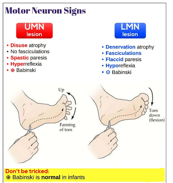
Question 109:
Which of the following is a unique characteristic of the cerebellum?
- Folia
- Betz cells
- Gyrus
- Fissures
- Pyramidal cells
Answer: A) Folia
Explanation: The cerebellum is characterized by the presence of folia, which are narrow, leaf-like gyri that create its highly folded surface. The folia increase the surface area of the cerebellum, allowing for more neurons and thus greater processing power for coordination and balance functions.
Option A is incorrect because Betz cells are large pyramidal neurons found in the primary motor cortex, not the cerebellum.
Option C is incorrect because gyrus (plural: gyri) are ridges found on the cerebral cortex.
Option D is incorrect because fissures are deep grooves that separate different regions of the brain, found throughout the brain, including the cerebellum.
Option E is incorrect because pyramidal cells are a type of neuron found in the cerebral cortex, particularly in areas involved in motor control and cognitive functions.
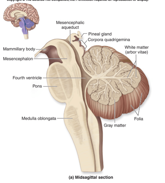
Question 110:
Which of the following is the main output of the cerebellum to the thalamus?
- 4th ventricle
- Superior peduncle
- Middle peduncle
- Inferior peduncle
- Folia
Answer: B) Superior peduncle
Explanation: The main output pathway of the cerebellum to the thalamus is through the superior cerebellar peduncle. This structure carries efferent fibres from the cerebellum to various parts of the brain, including the thalamus, which then relays the information to the cerebral cortex for motor control and coordination. This tract decussates.
Option A is incorrect because the 4th ventricle is a cavity within the brainstem, not involved in the direct output pathways.
Option C is incorrect because the middle peduncle primarily carries afferent fibres (input) from the cerebral cortex into the cerebellum. This tract decussates too.
Option D is incorrect because the inferior peduncle carries both afferent fibres (input) from the spinal cord to the cerebellum however this tract does not decussate.
Option E is incorrect because folia are the narrow, leaf-like gyri on the surface of the cerebellum, involved in increasing its surface area, not in direct neural pathways.
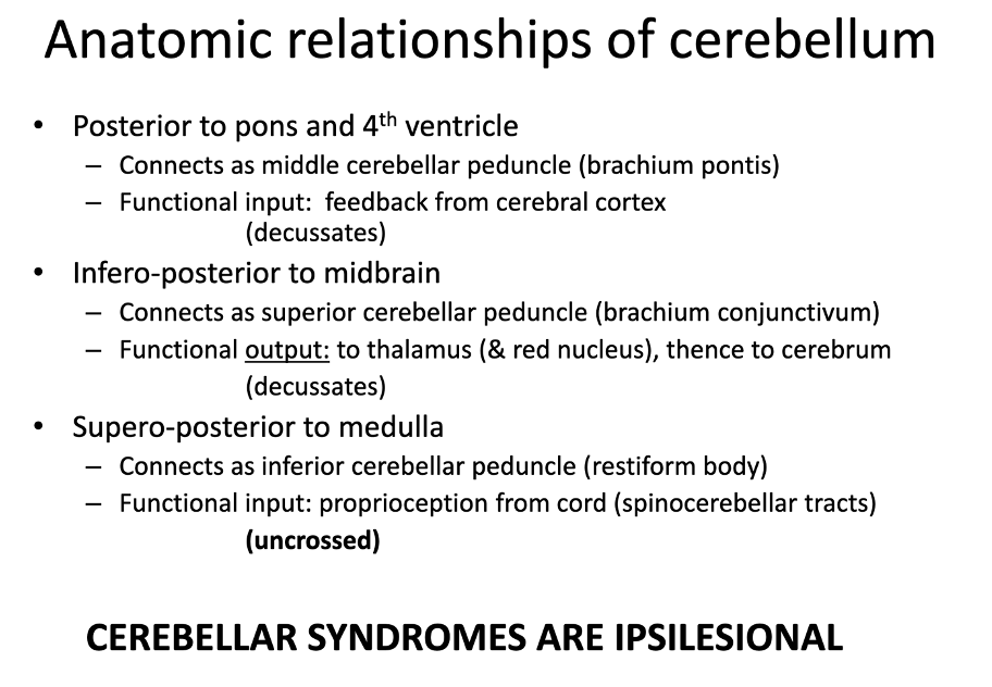
Question 111:
Which of the following is the main cerebellar nuclei found in the cerebrocerebellum?
- Dentate nucleus
- Interposed nucleus
- Globose nucleus
- Vestibular nucleus
- Fastigial nucleus
Answer: A) Dentate nucleus
Explanation: The main cerebellar nucleus found in the cerebrocerebellum is the dentate nucleus. The dentate nucleus is the largest and most lateral of the cerebellar nuclei and is primarily involved in the planning, initiation, and control of voluntary movements. It receives input from the lateral hemispheres of the cerebellum and projects to the thalamus and other brain regions to coordinate motor activity.
Option B is incorrect because the interposed nucleus, consisting of the globose and emboliform nuclei, is associated with the spinocerebellum and is involved in regulating motor execution.
Option C is incorrect because the globose nucleus, part of the interposed nucleus, also has a role in the spinocerebellum.
Option D is incorrect because the vestibular nucleus is involved with the vestibulocerebellum, which helps in maintaining balance and controlling eye movements.
Option E is incorrect because the fastigial nucleus is associated with the spinocerebellum and plays a role in posture and balance.

Question 112:
Which of the following is the deepest layer of the cerebellum?
- Purkinje layer
- Molecular layer
- Granule layer
- Pyramidal layer
- Folia
Answer: C) Granule layer
Explanation: The deepest layer of the cerebellum is the granule layer. This layer is located beneath the Purkinje layer and the molecular layer. It contains numerous small granule cells, which are the most abundant type of neuron in the brain. These granule cells receive input from mossy fibres and project their axons into the molecular layer, where they form parallel fibres that synapse with Purkinje cells. Unlike the neocortex of the cerebrum, the cerebellar cortex is uniform (i.e., the thickness of layers & types of cells is the same across the cerebellum). Whereas the neocortex differs in such a way that the predominant layer in the primary motor cortex is layer V, internal pyramidal layer, whilst the predominant layer in the primary somatosensory cortex is layer IV, internal granular layer.
Option A is incorrect because the Purkinje layer, situated above the granule layer, contains the cell bodies of Purkinje cells, which are large neurons that play a crucial role in motor coordination.
Option B is incorrect because the molecular layer is the outermost layer of the cerebellum, containing the dendrites of Purkinje cells, axons of granule cells, and various types of interneurons.
Option D is incorrect because the pyramidal layer is not a part of the cerebellar cortex but rather part of the cerebral cortex, specifically the primary motor cortex.
Option E is incorrect because folia are folds or ridges on the surface of the cerebellum that increase its surface area but are not considered a layer of the cerebellum.
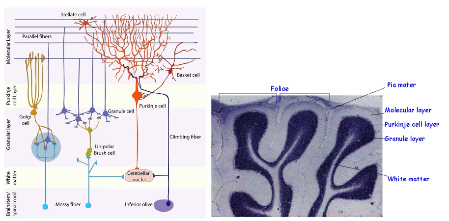
Question 113:
Climbing fibres that stimulate purkinje cells in the cerebellum project from which structure?
- Vestibular nucleus
- Olives of the medulla
- Pyramids of the medulla
- Inferior olivary nucleus
- Superior olivary nucleus
Answer: D) Inferior olivary nucleus
Explanation: Climbing fibers that stimulate Purkinje cells in the cerebellum originate from the inferior olivary nucleus. This nucleus, located in the medulla oblongata, sends climbing fibers that make direct excitatory synapses with Purkinje cells. These fibers are crucial for motor coordination and cerebellar learning. They play a significant role in error correction & only fire when an ‘error’ is detected such as an uncoordinated movement. This process is involved in a phenomenon called long-term depression (LTD), which refers to the weakening of synaptic connections between Purkinje cells and their granule cell inputs in response to climbing fibre activity. LTD is important for adjusting and fine-tuning motor commands and learning new motor skills, but it’s not the firing of climbing fibers that directly causes LTD; rather, the climbing fibers are involved in modulating the strength of synaptic connections through LTD.
- Role of Climbing Fibers: Climbing fibers from the inferior olivary nucleus send strong excitatory signals to Purkinje cells in the cerebellum. When these climbing fibers are active, they produce a large, powerful signal in Purkinje cells.
- Error Detection: When there’s a discrepancy between the expected and actual motor outcome (for example, if a movement doesn’t go as planned), climbing fibers detect this error.
- Induction of LTD: The strong activation of Purkinje cells by climbing fibers triggers LTD at the synapses between granule cells and Purkinje cells. Essentially, LTD reduces the strength of these synaptic connections.
- Purpose of LTD: By weakening these connections, the cerebellum can adjust and fine-tune motor commands for better accuracy in future movements. This adjustment helps improve motor control and learning by reducing the influence of erroneous signals and enhancing the overall precision of movement.
Option A is incorrect because the vestibular nucleus, while important for balance and spatial orientation, does not project climbing fibers to Purkinje cells. Instead, it sends input to the cerebellum through other pathways.
Option C is incorrect because the pyramids of the medulla are associated with the corticospinal tract, which transmits motor commands from the brain to the spinal cord.
Option E is incorrect because the superior olivary nucleus is involved in auditory processing and sound localisation.
Question 114:
Axons of which cell is the main output of the cerebellum?
- Mossy fibres
- Basket cells
- Granule cells
- Stellate cells
- Purkinje cells
Answer: E) Purkinje cell
Explanation: The main output of the cerebellum is carried by the axons of Purkinje cells. Purkinje cells receive signal from parallel fibres (axons of granule cells) & from climbing fibres. Purkinje cells are located in the Purkinje layer of the cerebellar cortex and always send inhibitory signals to the deep cerebellar nuclei. These nuclei then relay information to various motor and cognitive centres in the brain to coordinate and regulate movement.
Option A is incorrect because mossy fibbers are input pathways that carry information from all around the body (except from the inferior olivary nucleus) to granule cells within the cerebellum.
Option B is incorrect because basket cells are interneurons in the cerebellar cortex that modulate Purkinje cell activity but do not serve as the main output.
Option C is incorrect because granule cells are involved in processing input from mossy fibers and project their axons as parallel fibers to Purkinje cells.
Option D is incorrect because stellate cells are another type of interneuron in the cerebellar cortex that influences Purkinje cells but does not contribute to the main output of the cerebellum.
Question 115:
How many climbing fibres directly synapse with a single purkinje cell?
- 0
- 1
- 10
- 100
- 1000
Answer: B) 1
Explanation: A single Purkinje cell in the cerebellum is directly synapsed by 1 climbing fibre. Each Purkinje cell receives input from only one climbing fibre, although that climbing fibre can make multiple synaptic connections with the Purkinje cell.
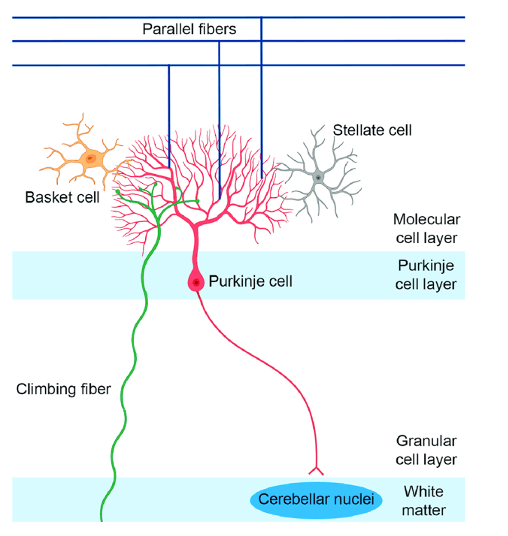
Question 116:
Mossy fibres synapse with which of the following cells?
- Granule cells & golgi cells
- Basket cells
- Cerebellar nuclei
- Purkinje cells
- Stellate cells
Answer: A) Granule & golgi cells
Explanation: Mossy fibers synapse with granule cells and Golgi cells in the cerebellum. Mossy fibers transmit information to granule cells, which then send their axons as parallel fibers to Purkinje cells. Mossy fibers also have an indirect effect on Golgi cells, which provide inhibitory feedback to granule cells.
Option B is incorrect because basket cells are interneurons that influence Purkinje cell activity but do not receive direct input from mossy fibres.
Option C is incorrect because the cerebellar nuclei receive inhibitory input from Purkinje cells rather than direct synapses from mossy fibres.
Option D is incorrect because purkinje cells are not directly synapsed by mossy fibres; they receive input from the parallel fibers of granule cells.
Option E is incorrect because stellate cells are also interneurons that modulate Purkinje cells but do not receive direct input from mossy fibers.
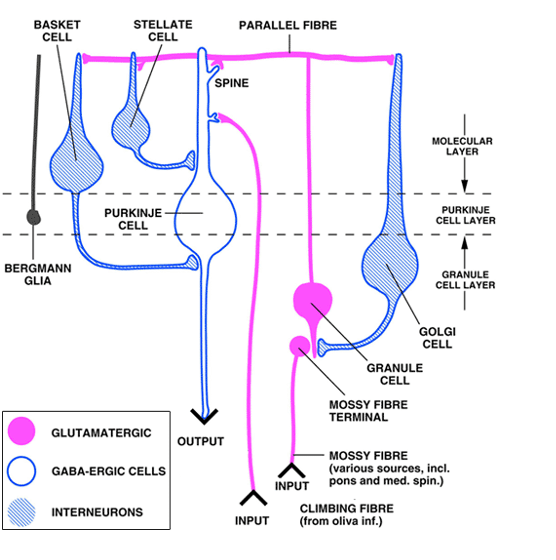
Question 117:
Where does all input to the cerebellum come from (except those from the inferior olivary nucleus)?
- Climbing fibres
- Medulla oblongata
- Mossy fibres
- Purkinje cells
- Granule cells
Answer: C) Mossy fibres
Explanation: All input to the cerebellum, except those from the inferior olivary nucleus, comes from mossy fibres. Mossy fibres carry information from various sources, including the spinal cord, brainstem, and other parts of the brain, to the cerebellum. They provide excitatory input to granule cells, which then influence Purkinje cells via their parallel fibres. Purkinje cells then inhibit deep cerebellar nuclei. Output of purkinje cells is always inhibitory.
Option A is incorrect because climbing fibres come from the inferior olivary nucleus & only fire when there is an ‘error’ e.g. an uncoordinated movement.
Option B is incorrect because the medulla oblongata is a structure that contains the inferior olivary nucleus but is not itself the source of all cerebellar input.
Option D is incorrect because purkinje cells are output neurons of the cerebellum.
Option E is incorrect because granule cells receive input from mossy fibers and send their axons as parallel fibers to Purkinje cells.
Question 118:
Damage to the cerebellum results in which of the following disorders?
- Ballismus
- Bradykinesia
- Dyskinesis
- Ataxia
- Apraxia
Answer: D) Ataxia
Explanation: Damage to the cerebellum results in ataxia. Ataxia is characterised by a lack of coordination and balance, affecting the precision and timing of movements. This occurs because the cerebellum is essential for coordinating voluntary movements and maintaining equilibrium.
Option A is incorrect because ballismus is a movement disorder involving violent, flinging movements, typically associated with lesions in the subthalamic nucleus of the basal ganglia, not the cerebellum.
Option B is incorrect because bradykinesia, or slowness of movement, is commonly associated with Parkinson’s disease and involves dysfunction of the basal ganglia.
Option C is incorrect because dyskinesis refers to abnormal & excessive involuntary movements associated with Huntington’s disease often due to damage in the basal ganglia.
Option E is incorrect because apraxia is a disorder involving difficulty in performing purposeful movements despite having the physical capability, usually related to cortical damage rather than cerebellar damage.
Question 119:
Which structures make up the neostriatum of the basal ganglia?
- Caudate & putamen
- Caudate & globus pallidus
- Caudate & nucleus accumbens
- Putamen & globus pallidus
- Putamen & substantia nigra
Answer: A) Caudate & putamen
Explanation: The structures that make up the neostriatum of the basal ganglia are the caudate and putamen. The neostriatum, also known as the striatum, is a major component of the basal ganglia involved in processing motor and cognitive information.
Question 120:
Which of the following structures is not a part of the basal ganglia?
- Substantia nigra
- Subthalamic nucleus
- Thalamus
- Nucleus accumbens
- Globus pallidus externus
Answer: C) Thalamus
Explanation: The structure that is not part of the basal ganglia is the thalamus. The thalamus is a major relay station in the brain that processes and transmits sensory and motor signals to the cerebral cortex but is not considered part of the basal ganglia.
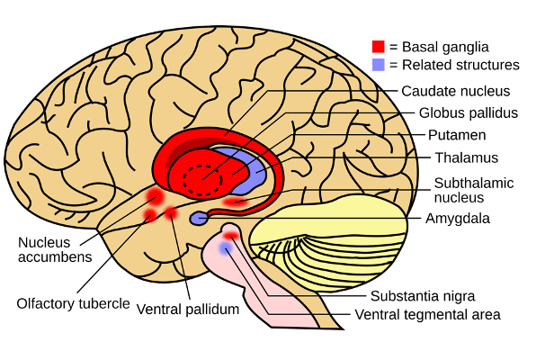
Question 121:
Which neurotransmitter does the substantia nigra release?
- Glutamate
- GABA
- Aspartate
- Serotonin
- Dopamine
Answer: E) Dopamine
Explanation: The substantia nigra releases dopamine specifically from an area called the pars compacta. Dopamine is a key neurotransmitter involved in regulating movement and reward pathways. Neurons in the substantia nigra project to other parts of the basal ganglia and use dopamine to modulate these circuits, which is crucial for smooth motor control.
Option A is incorrect because glutamate is an excitatory neurotransmitter found in various parts of the brain but not specifically released by the substantia nigra.
Option B is incorrect because GABA is an inhibitory neurotransmitter released by other neurons in the brain, including some within the basal ganglia, but it is not the primary neurotransmitter of the substantia nigra.
Option C is incorrect because aspartate, another excitatory neurotransmitter, is not primarily associated with the substantia nigra.
Option D is incorrect because serotonin is released by neurons in the raphe nuclei, not by the substantia nigra.
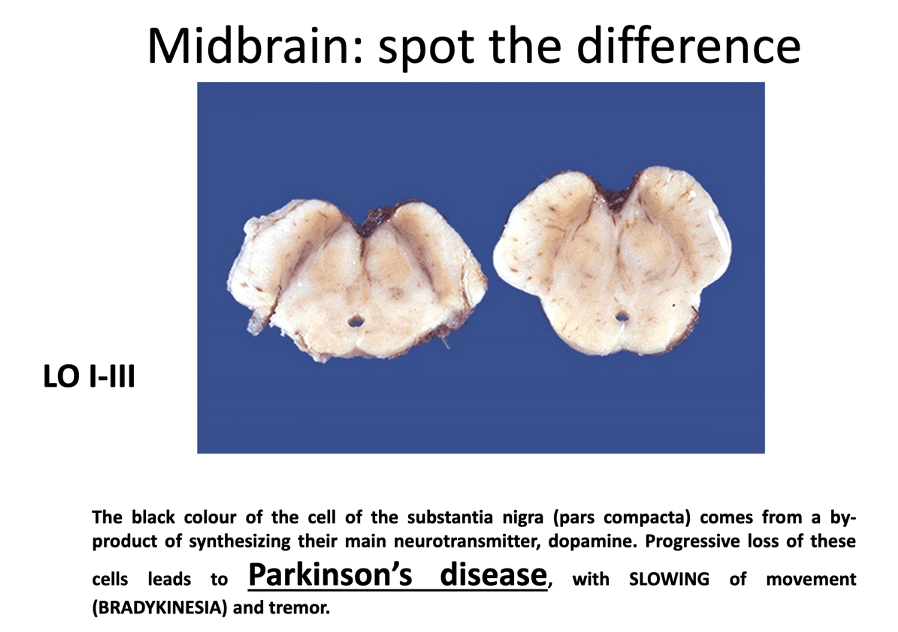
Question 122:
Which of the following receptors is associated with the indirect pathway of the basal ganglia?
- M3 – Muscarinic receptor
- Beta -2 adrenergic receptor
- D1
- D2
- D7
Answer: D) D2
Explanation: The receptor associated with the indirect pathway of the basal ganglia is the D2 receptor. The D2 receptors are located on neurons in the indirect pathway and play a role in modulating motor control by inhibiting movement. This pathway helps balance the overall output of the basal ganglia, influencing the regulation of voluntary movements.
Option A is incorrect because M3 muscarinic receptors are involved in parasympathetic nervous system functions.
Option B is incorrect because Beta-2 adrenergic receptors are involved in sympathetic nervous system responses.
Option C is incorrect because D1 receptors are associated with the direct pathway of the basal ganglia, which facilitates movement.
Option D is incorrect because D7 receptors are not specifically associated with the basal ganglia pathways involved in motor control.
Question 123:
What neurotransmitter does the striatum in the basal ganglia release in both the direct & indirect pathway, when activated by dopamine released from the substantia nigra?
- Dopamine
- GABA
- Glutamate
- Aspartate
- Glycine
Answer: B) GABA
Explanation: The striatum releases GABA, which is an inhibitory neurotransmitter, to modulate the activity of the basal ganglia circuits and influence motor control and other functions. GABA released from the striatum inhibits globus pallidus internus (in the direct pathway) & globus pallidus externus (in the indirect pathway).
Since globus pallidus internus is inhibited in the direct pathway, it can no longer release GABA to inhibit the thalamus. As a result, the thalamus secretes glutamate which activates & stimulates the motor cortex to produce movement.
In the indirect pathway, since globus pallidus externus is inhibited, it can no longer release GABA to inhibit the globus pallidus internus. As a result, since there is no inhibition on globus pallidus internus, GPi releases GABA which inhibits the thalamus. Since the thalamus is inhibited, it can no longer release glutamate to activate the motor cortex. This prevents any movement from occurring.
Note: All components of the basal ganglia release GABA when stimulated except for the thalamus & the motor cortex which release glutamate (excitatory)!
Question 124:
Which neurotransmitter does the thalamus release in the basal ganglia when it is not inhibited?
- Dopamine
- GABA
- Glutamate
- Aspartate
- Glycine
Answer: C) Glutamate
Explanation: When the thalamus is not inhibited in the basal ganglia, it releases glutamate. Glutamate is an excitatory neurotransmitter that promotes activity in the cortical areas by stimulating the neurons in the thalamus, which plays a role in modulating motor control and various sensory processes.


Question 125:
Which of the following components of the basal ganglia is involved with emotional processing?
- Putamen
- Caudate
- Globus pallidus
- Nucleus accumbens
- Subthalamic nucleus
Answer: D) Nucleus accumbens
Explanation: The nucleus accumbens is part of the ventral striatum and is integral to the brain’s reward system, motivation, and emotional regulation.
Option A is incorrect because putamen is mainly involved in motor control & processing.
Option B is incorrect because caudate is primarily associated with planning movements & behaviour.
Option C is incorrect because globus pallidus is involved in the output of the basal ganglia and primarily regulates movement, not emotional processes.
Option E is incorrect because the subthalamic nucleus is involved in motor control within the basal ganglia’s indirect pathway.
Question 126:
Damage to which of the following structures causes Huntington’s disease?
- Cerebellum
- Direct pathway of basal ganglia
- Direct & indirect pathway of the basal ganglia
- Indirect pathway of the basal ganglia
- Primary motor cortex
Answer: C) Direct & indirect pathway of basal ganglia
Explanation: Huntington’s disease is characterised by neurodegeneration that affects both pathways, leading to a decrease in the activity of the direct pathway and an increase in the activity of the indirect pathway. The indirect pathway degenerates earlier, leading to a loss of inhibition of the thalamus & therefore the primary motor causes which results in excessive unwanted movements called chorea. This disruption results in the motor and cognitive symptoms associated with the disease.
Option A is incorrect because damage to the cerebellum causes ataxia (uncoordinated movement).
Option B is incorrect because damage to the direct pathway of the basal ganglia alone causes Parkinson’s Disease (bradykinesia) with the main symptom being too little movement.
Option D is incorrect because damage solely to the indirect pathway does not fully explain the disorder, as Huntington’s disease impacts both pathways.
Option E is incorrect because the primary motor cortex is involved in executing movement but is not the primary site of damage in Huntington’s disease. Damage to the motor cortex causes apraxia which is difficulty in selecting the correct movement despite understanding the task/order.
Question 127:
Damage to which cortical area results in a lack of understanding of speech despite being able to produce fluent speech?
- Broca’s area
- Wernicke’s area
- Primary auditory cortex
- Hearing association area
- Left temporal lobe
Answer: B) Wernicke’s area
Explanation: Wernicke’s area, located in the left temporal lobe, is crucial for language comprehension. Individuals with damage to this area can speak fluently but their speech often lacks meaningful content, and they have difficulty understanding spoken language.
Option A is incorrect because Broca’s area is involved in speech production and damage to this area results in difficulty forming speech (non-fluent).
Option C is incorrect because the primary auditory cortex is responsible for processing auditory information but is not specifically involved in understanding speech.
Option D is incorrect because the hearing association area, or auditory association cortex, helps interpret sounds but does not specifically address the comprehension of speech.
Option E is incorrect because the left temporal lobe includes Wernicke’s area but is a broader region that also encompasses other functions beyond speech comprehension.
Note: Damage to Broca’s area causes expressive aphasia
Damage to Wernicke’s area causes receptive aphasia.
Second letter in Broca is ‘r’ & second letter in Wernicke is ‘e’. Swap them around.
Broca = expressive. Wernicke = receptive
Question 128:
Damage to the Papez circuit aka the limbic circuit causes which one of the following?
- Difficulty forming episodic memories
- Difficulty learning
- Difficulty forming procedural memories
- Difficulty forming semantic memories
- Difficulty recalling memories
Answer: A) Difficulty forming episodic memories
Explanation: Damage to the Papez circuit, also known as the limbic circuit, causes difficulty forming episodic memories. The Papez circuit is a network involved in the emotional and memory aspects of the limbic system, including the formation and retrieval of episodic memories, which are memories about personal experiences and events.
Option B is incorrect because difficulty learning is more associated with a broader range of cognitive processes and can involve multiple brain regions beyond the Papez circuit.
Option C is incorrect because difficulty forming procedural memories, which involve skills and habits, is not specifically linked to damage in the Papez circuit but rather to other brain areas like the basal ganglia and cerebellum.
Option D is incorrect because difficulty forming semantic memories, which correspond to general knowledge and facts, is related to damage in areas like the lateral temporal cortex.
Option E is incorrect because difficulty recalling memories is associated with general memory retrieval issues and may involve various brain regions, including but not limited to the Papez circuit.
Question 129:
Where does the Papez circuit begin?
- Fornix
- Thalamus
- Cingulum
- Hippocampus
- Parahippocampal gyrus
Answer: D) Hippocampus
Explanation: The Papez circuit begins in the hippocampus. The circuit starts with the hippocampus, which plays a crucial role in memory formation. From the hippocampus, information is relayed through the fornix to the mammillary bodies, and then to the anterior nuclei of the thalamus. It continues to the cingulate gyrus, across the cingulum and then back to the hippocampus via the parahippocampal gyrus, forming a loop crucial for emotional and memory processing.
A lesion anywhere in this circuit causes amnesia aka difficulty forming episodic memories.
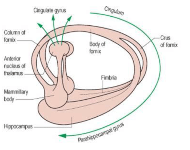
Question 130:
Which of the following brain structures is associated with fear & arousal?
- Parahippocampus
- Amygdala
- Limbic system
- Thalamus
- Epithalamus
Answer: B) Amygdala
Explanation: The amygdala is crucial for processing emotions, particularly fear, and plays a key role in the body’s response to arousal and stress.
Option A is incorrect because he parahippocampus is involved in memory and spatial navigation.
Option C is incorrect because the limbic system includes the amygdala and other structures involved in emotions and memory, but it is a broader network rather than a single structure specifically linked to fear and arousal.
Option D is incorrect because the thalamus acts as a relay station for sensory and motor signals but is not primarily associated with the processing of fear and arousal.
Option E is incorrect the epithalamus includes structures such as the pineal gland and is involved in regulating circadian rhythms by releasing melatonin.
